Skip to main page content
U.S. flag

An official website of the United States government

Dot gov

The . gov means it’s official. Federal government websites often end in . gov or . mil. Before sharing sensitive information, make sure you’re on a federal government site. VSports app下载.

Https

The site is secure V体育官网. The https:// ensures that you are connecting to the official website and that any information you provide is encrypted and transmitted securely. .

Comparative Study
. 2004 Jun 8;43(22):6987-94.
doi: 10.1021/bi049655t.

"V体育平台登录" Recognition of anionic phospholipid membranes by an antihemostatic protein from a blood-feeding insect

Affiliations
Comparative Study

Recognition of anionic phospholipid membranes by an antihemostatic protein from a blood-feeding insect

"V体育ios版" John F Andersen et al. Biochemistry. .

Abstract

The saliva of blood-feeding insects contains a variety of molecules having antihemostatic activity. Here, we describe nitrophorin 7 (NP7), a salivary protein that binds with high affinity to anionic phospholipid membranes VSports手机版. The protein is apparently targeted to the negatively charged surfaces of activated platelets and other cells, where it can serve as a vasodilator, antihistamine, platelet aggregation inhibitor, and anticoagulant. As with other members of the nitrophorin group, NP7 reversibly binds a molecule of NO and binds histamine with high affinity. The protein differs from other nitrophorins in that it binds to membranes containing phosphatidylserine. Sedimentation and surface plasmon resonance experiments, revealed two classes of phospholipid-binding sites having K(d) values of 4. 8 and 755 nM. NP7 inhibits prothrombin activation by blocking phospholipid binding sites for the prothrombinase complex on the surfaces of vesicles and activated platelets. As a NO complex, NP7 inhibits collagen and ADP-induced platelet aggregation and induces disaggregation of ADP-stimulated platelets by an NO-mediated mechanism. Molecular modeling of NP7 revealed a putative, positively charged membrane interaction surface comprised mainly of a helix lying outside of the lipocalin beta-barrel structure. .


Figures

Figure 1
Figure 1
Alignment of the NP7 amino acid sequence with NP2–4. The predicted signal peptide for NP7 is underlined. The sequence corresponding to the putative membrane-binding helix is boxed in blue in the NP7 sequence. The position of the K149A mutant is shown in green. The comparable sequences in NP2–4 are boxed in red.
Figure 2
Figure 2
NO and histamine complex formation with NP7 showing shift of the Soret maximum from 403 nm to 420 nm in the case of NO (-----) and 412 nm in the case of histamine (..-..-..-).
Figure 3
Figure 3
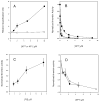
Binding of NPs with phospholipid vesicles as measured by sedimentation and SDS–PAGE. All assays were performed in 20 mM Tris-HCl, pH 7.5 containing the indicated concentrations of NaCl. In all cases, the phospholipid concentration was 0.5 mg/ml and the protein concentration was 3 μM. (A) Relationship of NP7 binding with buffer concentration of NaCl at various compositions of phospholipid. Filled circles: 100 % PS vesicles; filled squares: 3:1 PC:PS vesicles; open circles: 100 % PC vesicles. (B) Relationship of binding with NaCl concentration for NP1, NP2, NP3 and NP7, using 100 % PS vesicles. Filled circles: NP7; open circles: NP1, 2, 3. (C) Interaction of NP7 with a 3:1 PC:PS monolayer using surface plasmon resonance. Sensorgrams obtained with 30, 60, 200, and 600 nM NP7 are shown as solid lines. Sensorgram obtained with 220 nM NP3 is shown as a dashed line.
Figure 4
Figure 4
Anticoagulant activity of NP7. Each point represents the mean of three determinations. (A) Recalcification time of platelet-rich plasma (PRP) in the presence of NP7 and NP1. Citrated PRP was incubated with NP7 or NP1 as described in Materials and Methods, followed by addition of CaCl2 to a concentration of 5 mM. Filled circles, NP7; open circles, NP1. (B) Inhibition of reconstituted prothrombinase using various concentrations of 100% PS vesicles as a source of phospholipid. Thrombin generation was measured by the rate of hydrolysis of the chromogenic substrate S-2238. The relative thrombin activity is the rate of hydrolysis by thrombin formed in the presence of the indicated concentrations of NP7, divided by the rate formed in the absence of NP7. Filled circles, 0.5 μM PS; open circles, 1.0 μM PS; filled squares, 15 μM PS. (C) Relationship of prothrombinase inhibition with phospholipid concentration at a NP7 concentration of 230 nM, using 100% PS vesicles as a source of phospholipid. (D) Inhibition of prothrombinase activity using 3:1 PC:PS vesicles (1 μM) or collagen-activated platelets (1.5 × 10⁶ platelets in a final volume of 130 μl). Open circles: Prothrombinase activity (measured as rate of S-2238 hydrolysis) as a function of NP7 concentration in the presence of 3:1 PC:PS vesicles. Closed circles: Prothrombinase activity as a function of NP7 concentration with activated platelets serving as a source of phospholipid.
Figure 5
Figure 5
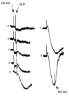
Inhibition of ADP-induced platelet aggregation by the NP7-NO complex as measured in an aggregometer. Washed platelets were treated with either NP7-NO complex or buffer (20 mM sodium acetate, pH 4.5 150 mM NaCl), then stimulated with ADP (3 μM). Concentrations of NP7-NO complex: a), 0 nM; b), 1 nM; c), 10 nM; d), 40 nM; e), 400 nM. Platelets were stimulated with ADP (3 μM), allowed to aggregate, then left untreated (f) or treated with NP7-NO complex (400 nM, g).
Figure 6
Figure 6
Molecular modeling of NP7. Models were constructed using the coordinates of the NP2 crystal structure (PDP accession number 1EUO (22)) with Swiss-Model. Heme was manually placed in model by superposition with NP2. Electrostatic potentials were calculated using DelPhi and a surface map was generated using GRASP. (A) Electrostatic potential surfaces of NP2 (left) and NP7 (right) showing the positively charged surface (blue) corresponding to the putative membrane interaction region. (B) Ribbon diagram of the NP7 model with lysine side chains (blue) highlighted on the external helix corresponding to the basic surface shown in Figure 6a.
Figure 6
Figure 6
Molecular modeling of NP7. Models were constructed using the coordinates of the NP2 crystal structure (PDP accession number 1EUO (22)) with Swiss-Model. Heme was manually placed in model by superposition with NP2. Electrostatic potentials were calculated using DelPhi and a surface map was generated using GRASP. (A) Electrostatic potential surfaces of NP2 (left) and NP7 (right) showing the positively charged surface (blue) corresponding to the putative membrane interaction region. (B) Ribbon diagram of the NP7 model with lysine side chains (blue) highlighted on the external helix corresponding to the basic surface shown in Figure 6a.
Figure 7
Figure 7
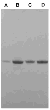
Sedimentation by ultracentrifugation of NP7 (K149A mutant) and NP7 (wild type) bound to 3:1 PC:PS vesicles at 200 mM NaCl. After sedimentation the supernatant was removed, and the pellet suspended in buffer and both were analyzed by SDS-PAGE with Coomassie blue staining. Lane A: NP7(K149A) pellet, Lane B: NP7 (K149A) supernatant, Lane C: NP7 (wild type) pellet, Lane D: NP7 (wild type) supernatant.

"VSports在线直播" References

    1. Nieswandt B, Watson SP. Platelet-collagen interaction: is GPVI the central receptor? Blood. 2003;102:449–461. - "VSports" PubMed
    1. Brass LF. More pieces of the platelet activation puzzle slide into place. J Clin Investig. 1999;104:1663–1665. - PMC - PubMed
    1. Coughlin SR. Protease-activated receptors in vascular biology. Thromb Haemost. 2001;86:298–307. - PubMed
    1. Sims PJ, Wiedmer T. Unraveling the mysteries of phospholipid scrambling. Thromb Haemost. 2001;86:266–275. - "VSports最新版本" PubMed
    1. Zwaal RF, Comfurius P, Bevers EM. Mechanism and function of changes in membrane-phospholipid asymmetry in platelets and erythrocytes. Biochem Soc Trans. 1993;21:248–253. - PubMed

"V体育官网" Publication types

MeSH terms (V体育2025版)