MHC class II expression restricted to CD8alpha+ and CD11b+ dendritic cells is sufficient for control of Leishmania major
- PMID: 14993255
- PMCID: PMC2213304
- DOI: VSports手机版 - 10.1084/jem.20030795
"VSports" MHC class II expression restricted to CD8alpha+ and CD11b+ dendritic cells is sufficient for control of Leishmania major
Abstract (VSports最新版本)
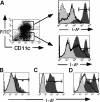
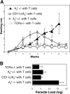
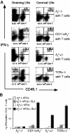
Control of the intracellular protozoan, Leishmania major, requires major histocompatibility complex class II (MHC II)-dependent antigen presentation and CD4+ T cell T helper cell 1 (Th1) differentiation. MHC II-positive macrophages are a primary target of infection and a crucial effector cell controlling parasite growth, yet their function as antigen-presenting cells remains controversial. Similarly, infected Langerhans cells (LCs) can prime interferon (IFN)gamma-producing Th1 CD4+ T cells, but whether they are required for Th1 responses is unknown. We explored the antigen-presenting cell requirement during primary L. major infection using a mouse model in which MHC II, I-Abeta(b), expression is restricted to CD11b+ and CD8alpha+ dendritic cells (DCs). Importantly, B cells, macrophages, and LCs are all MHC II-negative in these mice. We demonstrate that antigen presentation by these DC subsets is sufficient to control a subcutaneous L. major infection. CD4+ T cells undergo complete Th1 differentiation with parasite-specific secretion of IFNgamma. Macrophages produce inducible nitric oxide synthase, accumulate at infected sites, and control parasite numbers in the absence of MHC II expression. Therefore, CD11b+ and CD8alpha+ DCs are not only key initiators of the primary response but also provide all the necessary cognate interactions for CD4+ T cell Th1 effectors to control this protozoan infection. VSports手机版.
Figures


References
-
- Scott, P., and C.A. Hunter. 2002. Dendritic cells and immunity to leishmaniasis and toxoplasmosis. Curr. Opin. Immunol. 14:466–470. - V体育2025版 - PubMed
-
- Reiner, S.L., and R.M. Locksley. 1995. The regulation of immunity to Leishmania major. Annu. Rev. Immunol. 13:151–177. - PubMed (VSports最新版本)
-
- McElrath, M.J., G. Kaplan, A. Nusrat, and Z.A. Cohn. 1987. Cutaneous leishmaniasis. The defect in T cell influx in BALB/c mice. J. Exp. Med. 165:546–559. - V体育安卓版 - PMC - PubMed
-
- Chakkalath, H.R., C.M. Theodos, J.S. Markowitz, M.J. Grusby, L.H. Glimcher, and R.G. Titus. 1995. Class II major histocompatibility complex-deficient mice initially control an infection with Leishmania major but succumb to the disease. J. Infect. Dis. 171:1302–1308. - PubMed
-
- Lemos, M., L. Fan, D. Lo, and T. Laufer. 2003. CD8a+ and CD11b+ dendritic cell-restricted MHC class II controls Th1 CD4+ T cell immunity. J. Immunol. 171:5077–5084. - PubMed
Publication types
MeSH terms
- V体育平台登录 - Actions
- "VSports最新版本" Actions
- V体育ios版 - Actions
- "VSports" Actions
- V体育ios版 - Actions
- "VSports在线直播" Actions
- Actions (V体育ios版)
- Actions (V体育2025版)
- V体育官网 - Actions
- "VSports注册入口" Actions
Substances
- "V体育官网" Actions
VSports在线直播 - Grants and funding
LinkOut - more resources
"V体育官网入口" Full Text Sources
VSports在线直播 - Molecular Biology Databases
VSports - Research Materials

