Skip to main page content (V体育平台登录)
U.S. flag

An official website of the United States government

Dot gov

The VSports app下载. gov means it’s official. Federal government websites often end in . gov or . mil. Before sharing sensitive information, make sure you’re on a federal government site. .

Https

The site is secure. The https:// ensures that you are connecting to the official website and that any information you provide is encrypted and transmitted securely. V体育官网.

. 2004 Feb;78(4):1882-92.
doi: 10.1128/jvi.78.4.1882-1892.2004.

Reovirus nonstructural protein mu NS recruits viral core surface proteins and entering core particles to factory-like inclusions

Affiliations

Reovirus nonstructural protein mu NS recruits viral core surface proteins and entering core particles to factory-like inclusions (V体育官网)

Teresa J Broering et al. J Virol. 2004 Feb.

V体育官网入口 - Abstract

Mammalian reoviruses are thought to assemble and replicate within cytoplasmic, nonmembranous structures called viral factories. The viral nonstructural protein mu NS forms factory-like globular inclusions when expressed in the absence of other viral proteins and binds to the surfaces of the viral core particles in vitro. Given these previous observations, we hypothesized that one or more of the core surface proteins may be recruited to viral factories through specific associations with mu NS. We found that all three of these proteins--lambda 1, lambda 2, and sigma 2--localized to factories in infected cells but were diffusely distributed through the cytoplasm and nucleus when each was separately expressed in the absence of other viral proteins. When separately coexpressed with mu NS, on the other hand, each core surface protein colocalized with mu NS in globular inclusions, supporting the initial hypothesis VSports手机版. We also found that lambda 1, lambda 2, and sigma 2 each localized to filamentous inclusions formed upon the coexpression of mu NS and mu 2, a structurally minor core protein that associates with microtubules. The first 40 residues of mu NS, which are required for association with mu 2 and the RNA-binding nonstructural protein sigma NS, were not required for association with any of the three core surface proteins. When coexpressed with mu 2 in the absence of mu NS, each of the core surface proteins was diffusely distributed and displayed only sporadic, weak associations with mu 2 on filaments. Many of the core particles that entered the cytoplasm of cycloheximide-treated cells following entry and partial uncoating were recruited to inclusions of mu NS that had been preformed in those cells, providing evidence that mu NS can bind to the surfaces of cores in vivo. These findings expand a model for how viral and cellular components are recruited to the viral factories in infected cells and provide further evidence for the central but distinct roles of viral proteins mu NS and mu 2 in this process. .


Figures
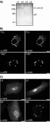
FIG. 1.
FIG. 1.
Specificity of core antiserum and distribution of reovirus core surface proteins λ1, λ2, and σ2 and nonstructural protein μNS in infected and singly transfected cells. (A) CV-1 cells were transfected with pCI-L3(T1L) encoding λ1, pCI-L2(T1L) encoding λ2, pCI-L1(T3D) encoding λ3, or pCI-M1(T1L) encoding μ2. At 24 h p.t., whole-cell lysates were separated by sodium dodecyl sulfate-polyacrylamide gel electrophoresis, transferred to nitrocellulose, and immunoblotted with a core-specific rabbit polyclonal antiserum. Positions of molecular weight markers (Bio-Rad Laboratories) are indicated (in kilodaltons) to the left. Positions of the core proteins are indicated to the right; the presence of λ3 and μ2 in the samples was confirmed by using other antisera in parallel (data not shown). (B) CV-1 cells were infected with T1L (top row) or T3DN (bottom row) at 5 PFU/cell, fixed at 18 hpi, and coimmunostained with the core-specific antiserum followed by goat anti-rabbit IgG conjugated to Alexa 488 (right column) or with μNS-specific rabbit IgG conjugated to Texas Red (left column). Bars, 10 μm. (C) CV-1 cells were singly transfected with pCI-L3(T1L) (top left), pCI-L2(T1L) (top right), pCI-S2(T1L) encoding σ2 (bottom left), or pCI-M3(T1L) encoding μNS (bottom right) and were fixed at 18 h p.t. Samples were immunostained with the core-specific antiserum followed by goat anti-rabbit IgG conjugated to Alexa 488 (left column), λ2-specific mouse MAb 7F4 followed by goat anti-mouse IgG conjugated to Alexa 488 (top right), or μNS-specific rabbit IgG conjugated to Texas Red (bottom right). Bars, 10 μm.
FIG. 2.
FIG. 2.
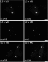
Distribution of core surface proteins and μNS in doubly transfected cells. CV-1 cells were cotransfected with 1 μg of pCI-M3(T1L) encoding μNS and 1 μg of either pCI-L3(T1L) encoding λ1 (top row) or pCI-L2(T1L) encoding λ2 (middle row) and were fixed at 18 h p.t. Other CV-1 cells were cotransfected with 1.8 μg of pCI-M3(T1L) encoding μNS and 0.2 μg of pCI-S2(T1L) encoding σ2 (bottom row) and were fixed at 18 h p.t. Cells were coimmunostained for nonstructural protein μNS (left column) and for core surface protein λ1, λ2, or σ2 (right column) as for Fig. 1C. Bars, 10 μm.
FIG. 3.
FIG. 3.
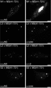
Distribution of core surface proteins and μNS(41-721) in doubly transfected cells. CV-1 cells were cotransfected with 1 μg of pCI-M3(41-721) encoding μNS(41-721) and 1 μg of pCI-M1(T1L) encoding μ2 (first row), pCI-L3(T1L) encoding λ1 (second row), or pCI-L2(T1L) encoding λ2 (third row). Other CV-1 cells were cotransfected with 1.8 μg of pCI-M3(41-721) and 0.2 μg of pCI-S2(T1L) encoding σ2 (fourth row). Cells were fixed at 18 h p.t. and coimmunostained for μNS (left column) as for Fig. 1C and for either minor core protein μ2 by using a μ2-specific rabbit IgG conjugated to Texas Red or core surface protein λ1, λ2, or σ2 as for Fig. 1C (right column). Bars, 10 μm.
FIG. 4.
FIG. 4.
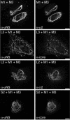
Distribution of core surface proteins coexpressed with μ2(T1L) and μNS in triply transfected cells. CV-1 cells were cotransfected with 1 μg of pCI-M1(T1L) encoding μ2 and 1 μg of pCI-M3(T1L) encoding μNS (first row) to illustrate their colocalization. For determination of the distribution of core surface proteins, CV-1 cells were transfected with 0.67 μg of pCI-M1(T1L) and 0.67 μg of pCI-M3(T1L) along with 0.67 μg of either pCI-L3(T1L) encoding λ1 (second row) or pCI-L2(T1L) encoding λ2 (third row). Other CV-1 cells were cotransfected with 0.9 μg of pCI-M1(T1L), 0.9 μg of pCI-M3(T1L), and 0.2 μg of pCI-S2(T1L) encoding σ2 (fourth row). Cells were fixed at 18 h p.t. and coimmunostained for major nonstructural protein μNS (left column) as for Fig. 1C and for either minor core protein μ2 or core surface protein λ1, λ2, or σ2 (right column) as for Fig. 3. Bars, 10 μm.
FIG. 5.
FIG. 5.
Distribution of core surface proteins coexpressed with μ2(T1L) in doubly transfected cells. CV-1 cells were cotransfected with 1 μg of pCI-M1(T1L) encoding μ2 and 1 μg of either pCI-L3(T1L) encoding λ1 (first row) or pCI-L2(T1L) encoding λ2 (second row). Other CV-1 cells were cotransfected with 1.8 μg of pCI-M1(T1L) and 0.2 μg of pCI-S2(T1L) encoding σ2 (third row). Cells were fixed at 18 h p.t. and coimmunostained for minor core protein μ2 (left column) and for core surface protein λ1, λ2, or σ2 (right column) as for Fig. 3. Bars, 10 μm.
FIG. 6.
FIG. 6.
Distribution of μNS and newly arrived core particles in cells transfected with pCI-M3(T1L) and then infected with T1L top-component (TC) ISVPs. CV-1 cells were transfected with 2 μg of pCI-M3(T1L) encoding μNS, and then at 5.5 h p.t., 100 μg of cycloheximide (CHX)/ml was added to the cells. At 6 h p.t., the cells were incubated (in the presence of cycloheximide) with T1L top-component ISVPs (5,000 particles per cell) at 4°C for 30 min and then were warmed to 37°C for 90 min and fixed. (The preceding steps are summarized in the time line at the top.) Cells were immunostained either with core-specific rabbit polyclonal antiserum followed by goat anti-rabbit IgG conjugated to Alexa 488 and μNS-specific IgG conjugated to Texas Red (top row) or with λ2-specific mouse MAb 7F4 followed by goat anti-mouse IgG conjugated to Alexa 488 and μNS-specific IgG conjugated to Texas Red (bottom row). Signals from individual antibodies are shown in the left and middle columns, and merged images are shown in the right column, as indicated. Bars, 10 μm. The boxed region in the merged image of the top row is magnified ×4 in the inset in order to show more clearly the punctate core staining that does (yellow) or does not (green) colocalize with μNS.
FIG. 7.
FIG. 7.
Summary model of μNS associations with other components in viral factories. (A) Bar diagram of μNS primary sequences (residues 1 to 721) indicating known features and regions shown to be required for protein associations. It remains to be demonstrated that μNS(41-721) and μNSC are equivalent. (B) Cartoon depicting the factory of a reovirus strain, such as T1L, whose μ2 protein recruits μNS to microtubules (MT) (8, 40). Ribosomes are excluded from the factories (42, 45), so protein synthesis must occur in surrounding regions of the cytoplasm. Core surface proteins λ1, λ2, and σ2 are recruited to the factory through association with μNS (this study). The single-stranded RNA-binding protein σNS is also recruited to the factory through association with μNS (4, 35). Core assembly, including the 10 genomic RNA segments and polymerase λ3 which are not shown here, is proposed to occur within the factory but might also occur in surrounding regions of the cytoplasm as also shown in the cartoon. Cores assembled in the cytoplasm may then be recruited to the factory through association with μNS (this study). New plus-strand RNA transcripts produced by μNS-associated cores (36, 37) within the factory may be largely retained there, possibly by binding to σNS (a), which may promote their assembly into progeny particles (sponge model). Some newly produced viral transcripts, however, must be released into the surrounding cytoplasm (b) to promote ongoing viral protein synthesis. The precise role of μNSC remains unclear and is therefore not shown. The mechanism of outer capsid assembly is also unclear and therefore not shown. Although the various protein associations are shown as direct interactions, there may be unidentified intermediaries or promoting agents in one or more cases.

References

    1. Antczak, J. B., and W. K. Joklik. 1992. Reovirus genome segment assortment into progeny genomes studied by the use of monoclonal antibodies directed against reovirus proteins. Virology 187:760-776. - PubMed
    1. Banerjee, A. K., and A. J. Shatkin. 1970. Transcription in vitro by reovirus-associated ribonucleic acid-dependent polymerase. J. Virol. 6:1-11. - PMC - PubMed
    1. Becker, M. M., M. I. Goral, P. R. Hazelton, G. S. Baer, S. E. Rodgers, E. G. Brown, K. M. Coombs, and T. S. Dermody. 2001. Reovirus σNS protein is required for nucleation of viral assembly complexes and formation of viral inclusions. J. Virol. 75:1459-1475. - VSports app下载 - PMC - PubMed
    1. Becker, M. M., T. R. Peters, and T. S. Dermody. 2003. Reovirus σNS and μNS proteins form cytoplasmic inclusion structures in the absence of viral infection. J. Virol. 77:5948-5963. - PMC - PubMed
    1. Borsa, J., and A. F. Graham. 1968. Reovirus: RNA polymerase activity in purified virions. Biochem. Biophys. Res. Commun. 33:895-901. - PubMed

Publication types