Skip to main page content
U.S. flag

An official website of the United States government

Dot gov

The . gov means it’s official. Federal government websites often end in . gov or . mil. Before sharing sensitive information, make sure you’re on a federal government site. VSports app下载.

Https

The site is secure. The https:// ensures that you are connecting to the official website and that any information you provide is encrypted and transmitted securely V体育官网. .

. 2004 Feb;74(2):239-52.
doi: 10.1086/381653. Epub 2004 Jan 19.

Ethylmalonic encephalopathy is caused by mutations in ETHE1, a gene encoding a mitochondrial matrix protein

Affiliations

Ethylmalonic encephalopathy is caused by mutations in ETHE1, a gene encoding a mitochondrial matrix protein

Valeria Tiranti et al. Am J Hum Genet. 2004 Feb.

Abstract

Ethylmalonic encephalopathy (EE) is a devastating infantile metabolic disorder affecting the brain, gastrointestinal tract, and peripheral vessels. High levels of ethylmalonic acid are detected in the body fluids, and cytochrome c oxidase activity is decreased in skeletal muscle. By use of a combination of homozygosity mapping, integration of physical and functional genomic data sets, and mutational screening, we identified GenBank D83198 as the gene responsible for EE. We also demonstrated that the D83198 protein product is targeted to mitochondria and internalized into the matrix after energy-dependent cleavage of a short leader peptide. The gene had previously been known as "HSCO" (for hepatoma subtracted clone one). However, given its role in EE, the name of the gene has been changed to "ETHE1 VSports手机版. " The severe consequences of its malfunctioning indicate an important role of the ETHE1 gene product in mitochondrial homeostasis and energy metabolism. .


"VSports注册入口" Figures
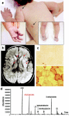
Figure  1
Figure 1
Clinical and biochemical features of EE. a, Skin areas with petechiae are indicated by arrows. The boxed pictures show acrocyanosis of hands and feet. b, On T2–fluid-attenuated-inversion-recovery (FLAIR) MRI images of a transverse section of the brain, symmetrical, patchy, high-intensity signals are present in the head of nucleus caudatus and in the putamen (arrows). c, Histochemical reaction to cytochrome c oxidase (COX) in skeletal muscle. The muscle of a patient with EE (top) shows a profound and diffuse reduction in COX reactivity, compared with an age-matched control biopsy (bottom). d, Gas-chromatographic profile of urinary organic acids in EE. The abnormal peak of EMA is indicated in red.
Figure  2
Figure 2
Pedigrees. The blackened symbols indicate the subjects with EE, and the unblackened symbols indicate the clinically healthy individuals. Beside each symbol are shown the individual haplotypes, covering a 3-cM region on chromosome 19q13. The markers from which the haplotypes were constructed and their genetic distances are shown in the boxed area against a chromosome-19 idiotype. The ETHE1 gene is flanked by markers D19S420 and D19S408.
Figure  3
Figure 3
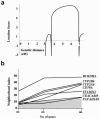
Genetic findings. a, Multipoint analysis of the EE locus for the families shown in figure 2. b, Integrative genomics strategy based on calculation of the mitochondrial neighborhood index. On the X-axis is shown the number of genes with expression profiles that were considered for the calculation of the index. The graph reports the values obtained for genes (UCSC annotation) from the 19q13 EE locus for which scores were equal to or higher than a threshold (gray area) corresponding to the score reported by Mootha et al. for the LRPPRC gene. YF13H12 (underlined) corresponds to a partial-length ETHE1 cDNA.
Figure  4
Figure 4
ETHE1 transcript and protein. a, Northern blot analysis of ETHE1 transcript on polyA+ mRNA extracted from different human tissues. B, brain; H, heart; M, skeletal muscle; C, colon; T, thymus; S, spleen; K, kidney; L, liver; I, small intestine; P, placenta; Lu, lung; Bl, blood. The 1,000-nt ETHE1 transcript is indicated by an arrow. b, Multiple alignments of the ETHE1 protein sequences in different species. Protein sequences were aligned using CLUSTALW. Amino acid substitutions are shown below the alignment, except for the I replacing the first M. A thick bar indicates the β-lactamase signature.
Figure  5
Figure 5
Characterization of ETHE1 gene and protein (ETHE1) in patients with EE. a, ETHE1 mutations identified in our patients. Mutations are indicated along the schematic representation of the ETHE1 cDNA. Exons are numbered in the cDNA scheme. The dark grey areas represent the 5′ and 3′ UTRs. The genomic organization of human ETHE1 is represented below the cDNA. b, Western blot analysis on cell lysates and isolated mitochondria from HeLa- and HeLaETHE1-derivative cell lines, using the anti-ETHE1C17 and anti-HA antibodies. The filter was treated with anti-ETHE1C17 and then stripped and rehybridized with the anti-HA antibody. The two antibodies show a virtually identical pattern in the HeLaETHE1 cell line, whereas ETHE1 protein CRM is barely detected by anti-ETHE1C17 antibody in naive, untransfected HeLa cells. c, Western blot analysis on fibroblast lysates was performed using anti-ETHE1C17; a monoclonal antibody specific for SDH-B, the 30-kDa subunit of succinate dehydrogenase, was used as a control. The same filter was first treated with anti-ETHE1C17 and then stripped and rehybridized with the anti-SDH antibody. C1 and C2 denote control fibroblasts. The upper and lower arrows indicate the precursor and mature ETHE1 polypeptides, respectively. Lanes A, B, E, I, J, K, and Q show fibroblasts from patients with EE. Each letter refers to the corresponding proband (and mutations) listed in table 2.
Figure  6
Figure 6
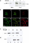
Mitochondrial localization of the ETHE1 protein. a, Mitochondrial import in vivo. Autoradiographs of 35S-radiolabeled HA-tagged ETHE1 protein, immunoprecipitated from HeLaETHE1 and Cos-7ETHE1 cell lysates. Arrows indicate the HA-tagged ETHE1 precursor protein, mature ETHE1 protein, and ETHE121, a shortened HA-tagged ETHE1 protein species lacking the first 20 amino acid residues on the N-terminus. Experiments were carried out in the presence (+) or absence (−) of valinomycin. b, Confocal immunofluorescence on Cos-7ETHE1 (panels 1–3 depict transient transfection) and HeLaETHE1 (panels 4–6 depict stable transfection). The green fluorescence in panels 1 and 4 corresponds to ETHE1HA protein–specific immunoreactivity. The red fluorescence in panels 3 and 6 corresponds to mitochondrial staining by MitoTracker, a mitochondrion-specific dye. The two immunofluorescence patterns largely overlapped, as shown by confocal merge images in panels 2 and 5. Magnification 40×. c, Proteinase K (PK)-protection assay by western blot analysis on freshly isolated mitochondria from human fibroblasts. Lanes 1–3 show intact isolated mitochondria treated with 0, 100, 250 μg/ml PK; lane 4 shows mitochondria treated with 0.5% Triton X-100 before digestion with 250 μg/ml PK. Anti-ETHE1C17 was used as the primary antibody. d, Suborganellar localization of the ETHE1 protein by western blot analysis on mitochondria isolated from human fibroblasts. Lane 1, cell lysate; lane 2, postmitochondrial supernatant (10,000 × g); lane 3, intact mitochondria; lane 4, mitochondrial membrane fraction; lane 5, mitochondrial soluble fraction. Anti-ETHE1C17 was used as the primary antibody.

References (VSports最新版本)

Electronic-Database Information

    1. Affymetrix, http://www.affymetrix.com/analysis/index.affx (VSports最新版本)
    1. CELERA, http://www.celera.com
    1. Center for Genome Research, http://www.genome.wi.mit.edu/mpg/isfc/ (for neighborhood index tables)
    1. CLUSTALW, "VSports在线直播" http://www.ebi.ac.uk/clustalw/
    1. Mitoprot-2, http://mips.gsf.de/cgi-bin/proj/medgen/mitofilter

References

    1. Aravind L (1999) An evolutionary classification of the metallo-β-lactamase fold proteins. In Silico Biol 1:69–91 - PubMed
    1. Attardi G, Schatz G (1988) Biogenesis of mitochondria. Annu Rev Cell Biol 4:289–333 - V体育官网 - PubMed
    1. Bito A, Haider M, Briza P, Strasser P, Breitenbach M (1999) Heterologous expression, purification, and kinetic comparison of the cytoplasmic and mitochondrial glyoxalase II enzymes, Glo2 and Glo4, from Saccharomyces cerevisiae. Protein Expr Purif 17:456–46410.1006/prep.1999.1151 - DOI - PubMed
    1. Bito A, Haider M, Hadler I, Breitenbach M (1997) Identification and phenotypic analysis of two glyoxalase II encoding genes from Saccharomyces cerevisiae, GLO2 and GLO4, and intracellular localization of the corresponding proteins. J Biol Chem 272:21509–2151910.1074/jbc.272.34.21509 - DOI - PubMed
    1. Burlina AB, Dionisi-Vici C, Bennet MJ, Gibson KM, Servidei S, Bertini E, Hale DE, Schimidt-Sommerfeld E, Sabetta G, Zacchello F, Rinaldo P (1994) A new syndrome with ethylmalonic aciduria and normal fatty acid oxidation in fibroblasts. J Pediatr 124:79–86 - PubMed

V体育平台登录 - Publication types

MeSH terms (VSports注册入口)

Substances (V体育安卓版)