V体育官网入口 - Skip to main page content
U.S. flag

An official website of the United States government

Dot gov

The . gov means it’s official. Federal government websites often end in VSports app下载. gov or . mil. Before sharing sensitive information, make sure you’re on a federal government site. .

Https

The site is secure. The https:// ensures that you are connecting to the official website and that any information you provide is encrypted and transmitted securely V体育官网. .

. 2003 Nov 9:3:30.
doi: 10.1186/1471-2407-3-30.

The histone deacetylase inhibitor Trichostatin A modulates CD4+ T cell responses

Affiliations

The histone deacetylase inhibitor Trichostatin A modulates CD4+ T cell responses

José Manuel Afonso Moreira et al. BMC Cancer. .

Abstract

Background: Histone deacetylase inhibitors (HDACIs) induce hyperacetylation of core histones modulating chromatin structure and affecting gene expression. These compounds are also able to induce growth arrest, cell differentiation, and apoptotic cell death of tumor cells in vitro as well as in vivo. Even though several genes modulated by HDAC inhibition have been identified, those genes clearly responsible for the biological effects of these drugs have remained elusive. We investigated the pharmacological effect of the HDACI and potential anti-cancer agent Trichostatin A (TSA) on primary T cells. VSports手机版.

Methods: To ascertain the effect of TSA on resting and activated T cells we used a model system where an enriched cell population consisting of primary T-cells was stimulated in vitro with immobilized anti-CD3/anti-CD28 antibodies whilst exposed to pharmacological concentrations of Trichostatin A V体育安卓版. .

Results: We found that this drug causes a rapid decline in cytokine expression, accumulation of cells in the G1 phase of the cell cycle, and induces apoptotic cell death. The mitochondrial respiratory chain (MRC) plays a critical role in the apoptotic response to TSA, as dissipation of mitochondrial membrane potential and reactive oxygen species (ROS) scavengers block TSA-induced T-cell death. Treatment of T cells with TSA results in the altered expression of a subset of genes involved in T cell responses, as assessed by microarray gene expression profiling V体育ios版. We also observed up- as well as down-regulation of various costimulatory/adhesion molecules, such as CD28 and CD154, important for T-cell function. .

Conclusions: Taken together, our findings indicate that HDAC inhibitors have an immunomodulatory potential that may contribute to the potency and specificity of these antineoplastic compounds and might be useful in the treatment of autoimmune disorders VSports最新版本. .


Figures

Figure 1
Figure 1
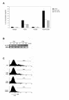
TSA induces apoptosis and cell cycle arrest in CD4+ T-cells. (A) CD4+ T-cells were treated for 24 hours with increasing concentrations of TSA (■). Cell death was determined by flow cytometric analysis of DNA content by PI staining of nuclei. Error bars show SD. (B) CD4+ T-cells were treated for 8 or 20 hours with various concentrations of TSA or DMSO alone as a negative control (-TSA). Cells were subsequently stained with Annexin V and PI. Inset in the bottom right quadrant of each plot are the percentage of cells that gated to the various quadrants (UL – upper left; UR – upper right; LL – lower left; and LR – lower right). The numbers for LR correspond to cells in early apoptosis (Annexin V positive and PI negative). (C) Effect of TSA on cell cycle progression of T cells. CD4+ T-cells were incubated for 24 h in the presence of medium containing IL-2 (10 U/ml), or anti-CD3 coupled beads (2:1 beads to cells ratio) after which either TSA (final concentration of 10 nM) or DMSO alone was added. After 20 hours the cells were analysed for cell cycle distribution by flow cytometry. Data are plotted as DNA content (propidium iodide fluorescence) vs relative live cell number (cells gated to R1). The total percentages of cells in the G0/G1 (region M1), S (region M2), and G2/M (region M3) phases of the cell cycle are shown. (D) Live TSA-treated cells (20 h with 10 nM TSA) were isolated by centrifugation in Ficoll Paque, washed extensively, labelled with CFSE and incubated in the presence of anti-CD3 coated beads (2:1 bead to cell ratio). Proliferation status of T cells was evaluated after 24 h (gray shaded histogram), 72 h (black open histogram), and 8 days (open gray histogram). Arrows indicate CFSE division peaks. For simplicity dead cells were excluded from the analysis. The data are representative of three independent experiments.
Figure 2
Figure 2
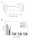
TSA generates ROS and activates the caspase-dependent cell death pathway. (A) Effect of cycloheximide and actinomycin-D on TSA-induced apoptosis. CD4+ T-cells were pre-treated with either CHX or act-D for 1 h after which TSA was added to the culture medium at a final concentration of 100 nM. After 20 h, levels of apoptosis were assessed by PI staining. (B) Western blots showing the effect of TSA on levels of caspase-3 and -10. CD4+ T-cells were cultured for 20 h in the presence of medium containing growing concentrations of TSA or DMSO alone (-TSA). After 20 hours the cells were harvested and cell extracts prepared as described in the Materials and Methods section. Western blots were prepared using one of the following antibodies: caspase-3 (H-277), or caspase-10 p20 (H-131) from Santa Cruz Biotechnology, Inc. (USA). Reversible staining of blots with Ponceau S was used as loading and blotting control. A typical blot is shown. (C) Effect of TSA on cell death receptor expression. Spleen cells from C57BL/6 mice were cultured for 48 h in the presence of soluble anti-CD3 antibody (0.5 μg/ml), whereafter CD4+ T-cells were isolated by magnetic cell separation as described (materials and methods section). Cells were subsequently stimulated with immobilized anti-CD3 mAb or cultured in the presence of IL-2 (10 U/ml), harvested after 20 hours in the presence of various concentrations of TSA, and the levels of CD95 and CD95L analysed by flow cytometry. Inset in the upper right quadrant of each plot are the percentage of cells that gated to the various quadrants (UL – upper left; UR – upper right; LL – lower left; and LR – lower right). Logic quadrants were placed such that the cells in a non-stimulated sample gate to the LL quadrant. Only live cells are included in the analysis. (D) TSA induces ROS generation. CD4+ T-cells were incubated for 24 h in the presence of medium containing IL-2 (10 U/ml), or anti-CD3 coupled beads (2:1 beads to cells ratio) after which either TSA (final concentration of 10 nM; bold black open histogram) or DMSO alone (black open histogram) was added. After 20 hours the cells were analysed for DHE and DCFDA fluorescence. (E) TSA-induced T-cell apoptosis can be inhibited by free radical scavengers and mitochondrial inhibitors. CD4+ T-cells were incubated for 24 h in the presence of medium containing IL-2 (10 U/ml), subsequently treated with SOD (2000 U/ml in the presence of 100 nM of digitonin), catalase (3000 U/ml), antimycin A (50 μM), or valinomycin (50 μM) for 1 h, after which TSA was added to the culture medium at growing concentrations. After 20 h, cells were harvested, and cell survival was determined by PI staining of nuclei. Shown are the mean percentage of survival; bars, SD. All data are representative of two independent experiments. A typical blot is shown.
Figure 3
Figure 3
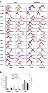
TSA represses IL-2 gene expression. Spleen cells from C57BL/6 mice were cultured for 48 h in the presence of soluble anti-CD3 antibody (0.5 μg/ml), whereafter CD4+ T-cells were isolated by magnetic cell separation as described in the materials and methods section. Cells were subsequently stimulated with the indicated immobilized antibodies and in the presence of the indicated inhibitors. (A) Inhibitors or DMSO alone (vehicle) were added to the medium – TSA (final concentration of 10 nM or 100 nM). After 8 hours the supernatant was harvested and IL-2 levels quantified by ELISA. (B) Purified CD4+ T-cells stimulated with immobilized anti-CD3 mAb were harvested after 4, 8, or 20 hours in the presence of 100 nM TSA, and levels of IL-2 mRNA quantified by relative RT-PCR as described (materials and methods section). Primers for 18S rRNA were used as control. (C) CD4+ T-cells stimulated with immobilized anti-CD3 mAb and cultured in growing concentrations of TSA were harvested after 8 hours and intracellular levels of IL-2 analysed by flow cytometry. Dead cells are excluded from the analysis. The data are representative of three independent experiments.
Figure 4
Figure 4
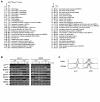
Addition of exogenous cytokines does not rescue TSA-treated cells. (A) Effect of IL-2 on cell survival. CD4+ T-cells were isolated by magnetic cell separation from C57BL/6 mouse splenocytes and cultured in medium containing 100 nM TSA (+TSA) or DMSO alone (-TSA) for 20 hours in the presence of growing concentrations of rmIL-2 (0–200 U/ml). Cell survival of TSA-treated cells is plotted in the right coordinates axis whereas that of non-treated cells is plotted in the left axis. (B) Effect of exogenous cytokine addition on cell survival. CD4+ T-cells were cultured in medium containing TSA at growing concentrations (+TSA) or DMSO alone (-TSA) and in the presence of various cytokines or stimuli (legend inset in graphic). PI staining of nuclei determined cell survival after 20 hours. Shown is the mean percentage of survival; bars represent SD. All data are representative of three independent experiments.
Figure 5
Figure 5
NFκB signalling is affected by TSA. (A) Effect of TSA on levels of nuclear NFκB. CD4+ T-cells were cultured with various concentrations of TSA (+TSA) or DMSO alone (-TSA) for 8 hours, whilst concomitantly stimulated with PMA+ionomycin (50 ng/ml and 1 μg/ml, respectively), or not stimulated (-PMA/ionomycin). Nuclear extracts were analysed for the presence of NFkB by western blot analysis using an antibody raised against NFκB p65 (C-20) (Santa Cruz Biotechnology, Inc. USA). (B) Effect of TSA on IκBα expression. CD4+ T-cells were pretreated with either 100 nM TSA (+TSA) or DMSO alone (-TSA) for 4 hours, whereafter phorbol-12-myristate-13-acetate (PMA; 50 ng/ml) and ionomycin (1 μg/ml), or vehicle (-PMA/ionomycin) was added to the medium and the cells were cultured for various periods of time. Cell lysates were analysed for IκBα expression by Western blot analysis using an anti-IκBα antibody from Cell Signaling Technology (CST, Beverly, MA). Reversible staining of blots with Ponceau S was used as loading and blotting control.
Figure 6
Figure 6
TSA modulates expression of cell surface molecules. (A) PBMCs from C57BL/6 mice were cultured in the presence of soluble anti-CD3 antibody (0.5 μg/ml) or IL-2 (10 U/ml) and exposed to 100 nM TSA for 4 hours (open black histogram) and 20 hours (open blue histogram), or to DMSO alone for 4 hours (open red histogram) or 20 hours (open brown histogram). Cells were harvested, extensively washed, and flow cytometric analysis of cell surface molecules performed as described in the materials and methods section. Dead cells are excluded from the analysis. (B) PBMCs from C57BL/6 mice were cultured in the presence of soluble anti-CD3 antibody (0.5 μg/ml) or IL-2 (10 U/ml) and exposed to 100 nM TSA or to DMSO alone for 20 hours. Cells were harvested, stained with a PE-conjugated anti-CD4 antibody and analysed by flow cytometry. PI staining of nuclei assessed cell viability. Shown are the mean percentage of survival; bars, SD. The data are representative of three independent experiments.
Figure 7
Figure 7
Transcriptional regulation of various genes by TSA (A) Gene expression profile of TSA-treated and non-treated CD4+ T-cells. Naïve CD4+ CD62L+ CD44low cells were isolated from C57BL/6 spleen cell preparations, cultured in the presence of IL-2 (10 U/ml) for 24 hours and Atlas Mouse cDNA Expression Arrays were used to analyse the differential gene expression caused by exposure of these cells to 100 nM TSA for 4 hours. Shown are all genes that varied in expression by greater than 2-fold in all experiments. Upward black arrows indicate upregulated genes, downward grey arrows denote downregulated genes. Data are based on two independent preparations of mRNA, for each experiment. Two membranes were used per sample. (B) Verification of several of the genes affected by TSA by semi-quantitative RT-PCR analysis. CD4+ T-cells were isolated by magnetic cell separation from pre-activated splenocytes (48 h in the presence of soluble anti-CD3 antibody). Cells were then stimulated with immobilized anti-CD3 mAb or cultured in the presence of IL-2 (10 U/ml), and harvested after 1, 4, or 20 hours in the presence of 100 nM TSA. Expression of 18S rRNA was used as normalizing control (C) Flow cytometric analysis of CD3ε and CD3 MC (molecular complex) expression in CD4+ T-cells exposed to TSA. CD4+ T-cells were cultured in the presence of immobilized anti-CD3 antibody (2:1 beads to cells ratio) or IL-2 (10 U/ml) and exposed to 100 nM TSA for 4 hours (open black histogram) and 20 hours (bold open black histogram), or to DMSO alone for 4 hours (open grey histogram) or 20 hours (open bold grey histogram). Data are representative of three independent experiments.

References

    1. Workman JL, Kingston RE. Alteration of nucleosome structure as a mechanism of transcriptional regulation. Annu Rev Biochem. 1998;67:545–579. doi: 10.1146/annurev.biochem.67.1.545. - DOI - PubMed
    1. Marmorstein R. Protein modules that manipulate histone tails for chromatin regulation. Nat Rev Mol Cell Biol. 2001;2:422–432. doi: 10.1038/35073047. - DOI - PubMed
    1. Spencer VA, Davie JR. Role of covalent modifications of histones in regulating gene expression. Gene. 1999;240:1–12. doi: 10.1016/S0378-1119(99)00405-9. - DOI - PubMed
    1. Rice JC, Allis CD. Histone methylation versus histone acetylation: new insights into epigenetic regulation. Curr Opin Cell Biol. 2001;13:263–273. doi: 10.1016/S0955-0674(00)00208-8. - DOI - PubMed
    1. Heinzel T, Lavinsky RM, Mullen TM, Söderstrom M, Laherty CD, Torchia J, Yang WM, Brard G, Ngo SD, Davie JR, Seto E, Eisenman RN, Rose DW, Glass CK, Rosenfeld MG. A complex containing N-CoR, mSin3 and histone deacetylase mediates transcriptional repression. Nature. 1997;387:43–48. doi: 10.1038/387043a0. - DOI - PubMed

MeSH terms