VSports手机版 - Skip to main page content
U.S. flag

An official website of the United States government

Dot gov

The . gov means it’s official. Federal government websites often end in VSports app下载. gov or . mil. Before sharing sensitive information, make sure you’re on a federal government site. .

Https

The site is secure V体育官网. The https:// ensures that you are connecting to the official website and that any information you provide is encrypted and transmitted securely. .

. 2003 Sep 16;100(19):10948-53.
doi: 10.1073/pnas.1833375100. Epub 2003 Sep 5.

Bacterial pathogens modulate an apoptosis differentiation program in human neutrophils

Affiliations

Bacterial pathogens modulate an apoptosis differentiation program in human neutrophils (VSports注册入口)

"VSports在线直播" Scott D Kobayashi et al. Proc Natl Acad Sci U S A. .

Abstract

Human polymorphonuclear leukocytes (PMNs or neutrophils) are essential to the innate immune response against bacterial pathogens. Recent evidence suggests that PMN apoptosis facilitates resolution of inflammation during bacterial infection. Although progress has been made toward understanding apoptosis in neutrophils, very little is known about transcriptional regulation of this process during bacterial infection. To gain insight into the molecular processes that facilitate resolution of infection, we measured global changes in PMN gene expression during phagocytosis of a diverse group of bacterial pathogens. Genes encoding key effectors of apoptosis were up-regulated, and receptors critical to innate immune function were down-regulated during apoptosis induced by phagocytosis of Burkholderia cepacia, Borrelia hermsii, Listeria monocytogenes, Staphylococcus aureus, and Streptococcus pyogenes. Importantly, we identified genes that comprise a common apoptosis differentiation program in human PMNs after phagocytosis of pathogenic bacteria. Unexpectedly, phagocytosis of Str. pyogenes induced changes in neutrophil gene expression not observed with other pathogens tested, including down-regulation of 21 genes involved in responses to IFN. Compared with other bacteria, PMN apoptosis was significantly accelerated by Str. pyogenes and was followed by necrosis VSports手机版. Thus, we hypothesize that there are two fundamental outcomes for the interaction of bacterial pathogens with neutrophils: (i) phagocytosis of bacteria induces an apoptosis differentiation program in human PMNs that contributes to resolution of bacterial infection, or (ii) phagocytosis of microorganisms such as Str. pyogenes alters the apoptosis differentiation program in neutrophils, resulting in pathogen survival and disease. .


Figures

Fig. 1.
Fig. 1.
Interaction of bacterial pathogens with human PMNs. (A) Phagocytosis of L. monocytogenes, Bkl. cepacia, Sta. aureus, and Str. pyogenes by human neutrophils. *, P < 0.001 vs. all bacteria; **, P ≤ 0.04 vs. all bacteria except heat-killed (Δ) Str. pyogenes. Results are the mean ± SD of three to five experiments. (B) Phagocytosis of Bor. hermsii.(Top) Number of PMNs with bound (black square) and/or ingested (green circle) Bor. hermsii. AI, association index; PI, phagocytic index. Micrographs (Right) illustrate ingestion and degradation of Bor. hermsii over time despite the appearance of extracellular staining (Extracellular, red). All Bor. hermsii, ingested, bound and extracellular (green). (Magnifications: ×400, Left; ≈×800, Right.) (C) PMN ROS production. The rate of ROS production for each pathogen is the mean of three separate experiments. ΔFL, change in fluorescence. (D) Killing of pathogens by human PMNs. At each time, PMNs were lysed and bacteria were plated on growth agar. Colonies were enumerated the next day, and percent bacteria killed was calculated as described in Supporting Methods. Results are the mean ± SD of three separate experiments. Bor. hermsii does not reliably form colony-forming units, and, thus, it was not possible to accurately assess PMN killing.
Fig. 2.
Fig. 2.
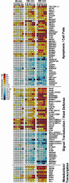
A common pathogen-induced transcription profile in human PMNs. Shown are genes differentially expressed during PMN phagocytosis. Scale bar at left indicates fold change. Results are the mean fold-induction or repression of genes from three separate microarray experiments using three blood donors with phagocytosis assays done on separate days. *, P ≤ 0.05 vs. unstimulated PMNs. LB, IgG and C3bi-coated latex beads (for reference); BH, Bor. hermsii; LM, L. monocytogenes; BC, Bkl. cepacia; SA, Sta. aureus; SP, Str. pyogenes.
Fig. 3.
Fig. 3.
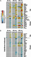
Str. pyogenes alters the PMN apoptosis differentiation program. (A) Str. pyogenes-specific modulation of apoptosis/cell fate-related genes in human PMNs. Symbols and scale bar are as described in the legend for Fig. 2. ΔSP, heat-killed Str. pyogenes. *, P ≤ 0.05 vs. unstimulated PMNs. (B) IFN-induced genes differentially expressed in PMNs after phagocytosis of Str. pyogenes.
Fig. 4.
Fig. 4.
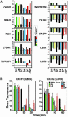
Confirmation of microarray results. (A) Genes (n = 10) identified as differentially transcribed by microarrays were analyzed by TaqMan real-time PCR. TNFRSF6 and TNFRSF10B are representative of genes modulated only by phagocytosis of Str. pyogenes. There was a strong positive correlation (r = 0.90) between TaqMan and microarray results, consistent with previous comparisons (10, 14). Each bar represents the average change in PMN gene expression from three individuals assayed in triplicate. Abbreviations are as in the Fig. 2 legend. (B) Flow cytometric analysis of IL-8 receptors. After phagocytosis of the indicated bacteria, expression of CXCR1 (Left) and CXCR2 (Right) was measured by flow cytometry. Results are the net mean fluorescence ± SD of two phagocytosis experiments.
Fig. 5.
Fig. 5.
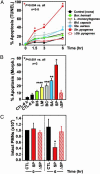
Bacterial pathogens induce PMN apoptosis. (A) PMN apoptosis was assessed with a modified terminal deoxynucleotidyltransferase-mediated dUTP nick end labeling assay after phagocytosis of pathogens. Red asterisk, P < 0.004 vs. unstimulated PMNs and cells stimulated with all other pathogens. Gray and blue asterisks, P < 0.03 vs. unstimulated PMNs. Results are the mean ± SD of three to five separate experiments. (B) PMN apoptosis was assessed by nuclear morphology (Morph.) after phagocytosis of pathogens. *, P ≤ 0.001 vs. unstimulated PMNs and cells stimulated with all other pathogens; **, P ≤ 0.003 vs. unstimulated PMNs and cells stimulated with Bor. hermsii, and heat-killed (Δ) Str. pyogenes. ***, P = 0.01 vs. unstimulated PMNs; ****, P = 0.04 vs. unstimulated PMNs. (C) Str. pyogenes induces PMN necrosis. PMN lysis was measured by microscopy at the indicated times after phagocytosis. *, P < 0.001 vs. unstimulated PMNs and heat-killed Str. pyogenes. Results are the mean ± SD of three to six separate experiments. Abbreviations are as in the Fig. 2 legend.

"V体育平台登录" References

    1. Kaplanski, G., Marin, V., Montero-Julian, F., Mantovani, A. & Farnarier, C. (2003) Trends Immunol. 24, 25–29. - PubMed
    1. Nauseef, W. M. & Clark, R. A. (2000) in Basic Principles in the Diagnosis and Management of Infectious Diseases, eds. Mandel, G. L., Bennett, J. E. & Dolin, R. (Churchill Livingstone, New York), Vol. 1, pp. 89–112.
    1. Lekstrom-Himes, J. A. & Gallin, J. I. (2000) N. Engl. J. Med. 343, 1703–1714. - PubMed
    1. Nathan, C. (2002) Nature 420, 846–852. - PubMed (VSports)
    1. Scaife, H., Woldehiwet, Z., Hart, C. A. & Edwards, S. W. (2003) Infect. Immun. 71, 1995–2001. - V体育官网入口 - PMC - PubMed