Skip to main page content
U.S. flag

An official website of the United States government

Dot gov

The . gov means it’s official. Federal government websites often end in . gov or . mil. Before sharing sensitive information, make sure you’re on a federal government site VSports app下载. .

Https

The site is secure V体育官网. The https:// ensures that you are connecting to the official website and that any information you provide is encrypted and transmitted securely. .

. 2003 Aug 19;100(17):9855-60.
doi: 10.1073/pnas.1733908100. Epub 2003 Aug 6.

VSports在线直播 - Targeted disruption of p185/Cul7 gene results in abnormal vascular morphogenesis

Affiliations

Targeted disruption of p185/Cul7 gene results in abnormal vascular morphogenesis (V体育ios版)

Takehiro Arai et al. Proc Natl Acad Sci U S A. .

Abstract

Cul1, a member of the cullin ubiquitin ligase family, forms a multiprotein complex known as SCF and plays an essential role in numerous cellular and biological activities. A Cul1 homologue, p185 (Cul7), has been isolated as an simian virus 40 large T antigen-binding protein. To understand the physiological role of p185, we generated mice lacking p185 VSports手机版. p185-/- embryos are runted and die immediately after birth because of respiratory distress. Dermal and hypodermal hemorrhage is detected in mutant embryos at late gestational stage. p185-/- placentas show defects in the differentiation of the trophoblast lineage with an abnormal vascular structure. We demonstrate that p185 forms an SCF-like complex with Skp1, Rbx1, Fbw6 (Fbx29), and FAP68 (FAP48, glomulin). FAP68 has recently been identified as a gene responsible for familial glomuvenous malformation. These results suggest that p185 forms a multiprotein complex and plays an important role in vascular morphogenesis. .


Figures
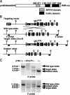
Fig. 1.
Fig. 1.
Targeted disruption of p185 gene. (A) Domain structure of human p185 predicted by National Center for Biotechnology Information Conserved Domain Search. The residue for the beginning and end of each domain is shown at the top. (B) Genomic organization of the amino-terminal region of mouse p185, targeting vector and mutant allele after homologous and Cre recombination. Exon 2 contains the translation start site. The filled triangle and open rectangle indicate loxP and Frt sites, respectively. D, DraI; B, BspHI; DT, diphtheria toxin; NEO, neomycin acetyltransferase. (C) Southern blot analysis of p185+/+, p185+/2loxP, and p185+/ mice and p185/embryo. DraI- and BspHI-digested genomic DNA was hybridized with 5′ and 3′ probes, respectively.
Fig. 2.
Fig. 2.
Neonatal death and growth retardation of p185/ embryo. (A) Genotype and phenotype of p185+/ intercrosses. Dead embryos are defined as necrotic or partially absorbed fetuses (indicated by *). %–/–, proportion of viable p185/embryos or pups at each stage; % weight, average of percentage weight of each p185/ embryo against the average weight of p185+/ and p185+/+ littermates. The weights of p185+/ and p185+/+ embryos were indistinguishable (data not shown). (B) Gross appearance of p185+/+ and p185/ embryos and placentas at indicated embryonic day. (Scale bar, 10 mm.) (C) Growth kinetics of MEFs. Passage-2 MEFs (1 × 10⁵) were plated in 60-mm dishes and counted every 24 h. •, p185+/+; □, p185+/; ▴, p185/. (D) The lung of E18.5 mutant embryo was unable to inflate, compared with a wild-type littermate. (×20.)
Fig. 3.
Fig. 3.
Histological abnormality of p185/ embryos and placentas. (A) Sagittal section of a mutant embryo showed dermal (arrowhead) and hypodermal hemorrhage in lower lip region. (×20.) (B) In situ hybridization with spongiotrophoblast-specific marker, Tpbp. The spongiotrophoblast (Sp) and decidual layers (Dec) were reduced in E18.5 mutant placenta compared with the wild-type littermate. Lt, labyrinthine. (Scale bar, 1 mm.) (C) Hematoxylin/eosin-stained E18.5 placenta reveals secondary trophoblast giant cells (arrowhead) were present in wild-type placenta, whereas they were virtually absent in p185/ placenta. Dilated maternal vessels (arrow) were detected in spongiotrophoblast layer of wild-type placenta, and only small vessels were detected in p185/ placenta. (×2.) (D) E12.5 mutant labyrinthine contains fewer maternal (red) and more fetal (blue) vessels than wild type. Fetal and maternal vessels are distinguished by the size and nucleation of red blood cells. (×20.) (E) The total area of fetal and maternal vessels in labyrinthine was measured by NIH image software. Twelve different fields (×100) for each genotype were randomly selected for analysis, and the average of percentage area in each field was calculated. Significant differences between p185/ and p185+/+ or p185+/ placenta were found in both fetal and maternal vessel areas. *, P < 0.05; **, P < 0.01.
Fig. 4.
Fig. 4.
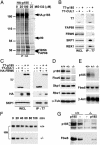
p185 associates with FBW6, FAP68, SKP1, and RBX1. (A) Identification of [35S]methionine-labeled proteins that specifically coimmunoprecipitate with HA-p185. U-2 OS cells stably expressing HA-p185 were treated with the indicated amount of MG132 and immunoprecipitated with anti-HA antibody (12CA5). Retained proteins were resolved by SDS/PAGE and visualized by autoradiography. Subsequently, 90-kDa and 58-kDa bands were cut out from the silver-stained gel and subjected to mass spectrometric analysis. The 58-kDa band contained two different proteins, FBW6 and FAP68. No meaningful peptide sequence was recovered from the 90-kDa band. (B) T7-p185 associates with endogenous FBW6 and FAP68 in addition to SKP1 and RBX1. Lysates from HeLa cells transfected with indicated constructs were immunoprecipitated with anti-T7 antibody and analyzed by immunoblotting with anti-FBW6, FAP68, SKP1, and RBX1 antibodies. (C) p185 binds to SKP1 in an FBW6-dependent manner. T7-p185 or T7-Cul1 were transfected into HeLa cells with HA-Fbw6. Immunoblotting was performed with indicated antibodies for cell extract and immunoprecipitates. (D) Expression of Fbw6 protein in p185/ MEFs. Immunoblotting was performed on whole cell lysates (100 μg per lane) prepared from p185+/+, p185+/, and p185/ MEFs at passage 2. The membranes were probed with anti-p185 and Fbw6 antibodies. (E) mRNA expression of Fbw6 in p185/ MEFs. Total mRNA (14 μg per lane) from p185+/+, p185+/, and p185/ MEFs at passage 2 were analyzed by using radiolabeled p185 and Fbw6 probes. (F) p185+/+ and p185/ MEFs stably expressing HA-tagged mouse Fbw6 were treated with 50 μg/ml cycloheximide and harvested every 20 min. The membrane was immunoblotted with anti-HA. Half-life of Fbw6 in p185/ and p185+/+ MEFs was 51 and 111 min, respectively. (G) Lysates from p185+/+ and p185/ MEFs that were transduced with a retroviral vector only (–) or containing wild-type mouse p185 were immunoblotted with p185 or Fbw6 antibodies.
Fig. 5.
Fig. 5.
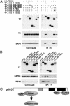
Overall structure of p185-based complex. (A) FBW6 binds to amino-terminal region of p185. HeLa cells were cotransfected with HA-FBW6 and six different p185 constructs. Lysates of HeLa cells were immunoprecipitated with anti-T7 and analyzed by anti-HA and SKP1 antibodies. (B) FAP68 binds to the carboxyl-terminal region of p185. Lysates from HeLa cells transfected with p185 deletion constructs were immunoprecipitated with anti-T7 and analyzed by immunoblotting with anti-FAP68 and RBX1 antibodies. (C) A model of p185-based complex.

"V体育官网" References

    1. Hershko, A. & Ciechanover, A. (1998) Annu. Rev. Biochem. 67, 425–479. - PubMed
    1. Deshaies, R. J. (1999) Annu. Rev. Cell Dev. Biol. 15, 435–467. - PubMed (V体育2025版)
    1. Zhang, H., Kobayashi, R., Galaktionov, K. & Beach, D. (1995) Cell 82, 915–925. - PubMed
    1. Koepp, D. M., Harper, J. W. & Elledge, S. J. (1999) Cell 97, 431–434. - "V体育ios版" PubMed
    1. Bodine, S. C., Latres, E., Baumhueter, S., Lai, V. K., Nunez, L., Clarke, B. A., Poueymirou, W. T., Panaro, F. J., Na, E., Dharmarajan, K., et al. (2001) Science 294, 1704–1708. - PubMed

Publication types

MeSH terms