VSports - Skip to main page content
U.S. flag

An official website of the United States government

Dot gov

The . gov means it’s official. Federal government websites often end in . gov or . mil VSports app下载. Before sharing sensitive information, make sure you’re on a federal government site. .

Https

The site is secure. The https:// ensures that you are connecting to the official website and that any information you provide is encrypted and transmitted securely V体育官网. .

. 2003 Jun 16;197(12):1709-19.
doi: 10.1084/jem.20030170.

Unraveling a revealing paradox: Why major histocompatibility complex I-signaled thymocytes "paradoxically" appear as CD4+8lo transitional cells during positive selection of CD8+ T cells

Affiliations

Unraveling a revealing paradox: Why major histocompatibility complex I-signaled thymocytes "paradoxically" appear as CD4+8lo transitional cells during positive selection of CD8+ T cells (VSports app下载)

Remy Bosselut et al. J Exp Med. .

Abstract

The mechanism by which T cell receptor specificity determines the outcome of the CD4/CD8 lineage decision in the thymus is not known. An important clue is the fact that major histocompatibility complex (MHC)-I-signaled thymocytes paradoxically appear as CD4+8lo transitional cells during their differentiation into CD8+ T cells. Lineage commitment is generally thought to occur at the CD4+8+ (double positive) stage of differentiation and to result in silencing of the opposite coreceptor gene. From this perspective, the appearance of MHC-I-signaled thymocytes as CD4+8lo cells would be due to effects on CD8 surface protein expression, not CD8 gene expression. But contrary to this perspective, this study demonstrates that MHC-I-signaled thymocytes appear as CD4+8lo cells because of transient down-regulation of CD8 gene expression, not because of changes in CD8 surface protein expression or distribution. This study also demonstrates that initial cessation of CD8 gene expression in MHC-I-signaled thymocytes is not necessarily indicative of commitment to the CD4+ T cell lineage, as such thymocytes retain the potential to differentiate into CD8+ T cells. These results challenge classical concepts of lineage commitment but fulfill predictions of the kinetic signaling model VSports手机版. .


Figures (VSports注册入口)

Figure 1.
Figure 1.
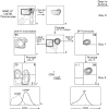
Alternative schematic representations of the mechanism by which MHC-I–signaled thymocytes differentiate into CD8+ T cells. Differentiation of MHC-I–signaled thymocytes is schematized as a two color plot of surface CD4 versus CD8 expression of MHC-IIo thymocytes. Labels outside the plot identify thymocyte subpopulations by surface levels of CD4/CD8 protein expression. Labels inside the cells indicate coreceptor gene expression. Arrows indicate proposed precursor/progeny relationships according to either classical lineage commitment models (A) or the kinetic signaling model (B). Cells in light gray are absent in MHC-IIo thymi but present in normal thymi. (A) Classical lineage commitment models (e.g., instruction, stochastic/selection, strength of signal, duration of signal) are all based on the central paradigm that lineage commitment occurs in DP thymocytes and results in silencing of the opposite coreceptor gene. As a result, all classical models require that MHC-I–signaled thymocytes appear as CD4+8lo transitional cells because of changes in CD8 protein expression, not CD8 gene expression. (B) The kinetic signaling model proposes that TCR-signaled DP thymocytes initially terminate CD8 gene expression and appear as CD4+8lo transitional cells as a result of diminished or absent CD8 gene expression. In the kinetic signaling model, TCR disengagement of MHC-I ligands occurs in CD4+8lo transitional cells because of diminished surface CD8 coreceptor levels and results in cessation of TCR signaling, reversal of coreceptor gene expression, and terminal differentiation into CD8+ T cells.
Figure 2.
Figure 2.
Differential expression of endogenous and transgenic CD8α expression on thymocytes signaled in vivo by intrathymic MHC-I complexes. Thymocytes from MHC-II0 mice (either with or without the CD8α transgene) were surface stained for TCRβ, CD4, CD8β, and either CD8α.2 (endogenous CD8α) or CD8α.1 (transgenic CD8α) expression and analyzed by four color flow cytometry. Shown on whole thymus populations from each mouse are CD4 versus CD8β and TCRβ expression (left panels). Because intrathymically signaled thymocytes up-regulate surface TCR expression and become TCRhi cells, we gated on TCRhi thymocytes to determine surface coreceptor expression on in vivo–signaled cells (right panels). CD4 versus CD8β expression on TCRhi thymocytes is shown, with CD8βlo cells identified by the blue rectangular gate and CD8βhi identified by the red rectangular gate. CD8α staining on CD8βlo (blue line) and CD8βhi (red line) TCRhi thymocytes was compared for surface expression of endogenous CD8α.2 and transgenic CD8α.1 molecules on the identical cells. Surface expression of endogenous CD8α.2 molecules was found to parallel that of CD8β, in that CD8α.2 expression was low on CD8βlo cells and high on CD8βhi TCRhi cells. In contrast, surface expression of transgenic CD8α.1 molecules did not parallel that of CD8β, in that CD8α.1 expression was identical on CD8βlo and CD8βhi TCRhi thymocytes.
Figure 3.
Figure 3.
Effect of intrathymic MHC-I signals on surface expression of full-length CD8α and tailless CD8α′ transgenic molecules. Thymocytes were obtained from MHC-IIo mice that expressed only endogenous CD8 molecules (both CD8α and CD8α′; top row), or from MHC-IIo CD8αo mice that either expressed only full-length CD8α (middle row) or tailless CD8α′ (bottom row) transgenic molecules. Thymocytes were subjected to the same four color flow cytometric analysis as in Fig. 2, with the exception that both endogenous and transgenic CD8α molecules were stained with a pan anti-CD8α mAb. In contrast to endogenous CD8α molecules that were expressed at significantly lower levels on CD8βlo (blue line) than CD8βhi (red line) TCRhi thymocytes, transgenic CD8α molecules were expressed at similar levels on CD8βlo and CD8βhi TCRhi thymocytes regardless of whether the transgene encoded full-length CD8α or tailless CD8α′ molecules.
Figure 4.
Figure 4.
Effect of intrathymic MHC-I signals on surface expression of CD8αα homodimeric complexes. Thymocytes were obtained from MHC-IIo CD8βo mice that expressed both endogenous CD8α.2 and transgenic CD8α.1 molecules as CD8αα complexes. Thymocytes were surface stained for TCRβ, CD4, CD8β, and either CD8α.2 (endogenous CD8α) or CD8α.1 (transgenic CD8α) expression and analyzed by four color flow cytometry. Shown on whole thymus populations are CD4 versus CD8β expression, CD4 versus CD8α.2 expression, and TCRβ expression (left panels). CD4 versus CD8α.2 expression on gated TCRhi thymocytes is shown and identifies CD8α.2lo cells (mostly CD4+ cells, blue rectangular gate) and CD8 α.2hi cells (mostly CD4 cells, red rectangular gate). CD8α.1 surface expression was found to be similar on CD8α.2lo (blue line) and CD8α.2hi (red line) TCRhi thymocytes (right panel).
Figure 5.
Figure 5.
Reexpression rates of endogenous and transgenic coreceptor proteins. (A) Thymocytes from MHC-IIo mice that carried the CD8α transgene and expressed both endogenous CD8α.2 and transgenic CD8α.1 molecules were stripped of all surface coreceptor proteins by treatment with extracellular pronase. The stripped thymocytes were cultured in single cell suspensions at 37°C for varying times, and assessed for reexpression of CD4, CD8α.2, and CD8α.1 by staining with anti-CD4 and allele-specific anti-CD8 mAbs. Displayed are histograms of CD4 versus CD8α.2 and CD4 versus CD8α.1, reexpression at different times after pronase stripping. Although CD4 reexpression occurred more quickly than CD8 reexpression, the kinetics of reexpression of endogenous CD8α.2 and transgenic CD8α.1 molecules were identical. (B) Thymocytes were obtained from MHC-IIo mice expressing only endogenous CD8α.2 molecules, and from MHC-IIo CD8αo mice that contained the CD8α transgene and therefore expressed only transgenic CD8α.1 molecules. Thymocytes were stripped of surface coreceptor proteins by treatment with extracellular pronase, cultured in single cell suspensions at 37°C for varying times, and assessed for reexpression of CD4 and CD8α by staining with anti-CD4 and a pan anti-CD8 mAb that recognizes both CD8α.1 and CD8α.2 isoforms. Line graphs show quantitative reexpression of CD4 and CD8 surface proteins relative to their maximum reexpression at 24 h, which was set equal to 100%. The kinetics of CD4 reexpression were identical on thymocytes from MHC-IIo and CD8α transgenic MHC-IIo CD8αo mice, but are displayed only for the former. Quantitation of surface reexpression versus time of CD4 (blue line), endogenous CD8α.2 (red line), and transgenic CD8α.1 (green line) are displayed.
Figure 5.
Figure 5.
Reexpression rates of endogenous and transgenic coreceptor proteins. (A) Thymocytes from MHC-IIo mice that carried the CD8α transgene and expressed both endogenous CD8α.2 and transgenic CD8α.1 molecules were stripped of all surface coreceptor proteins by treatment with extracellular pronase. The stripped thymocytes were cultured in single cell suspensions at 37°C for varying times, and assessed for reexpression of CD4, CD8α.2, and CD8α.1 by staining with anti-CD4 and allele-specific anti-CD8 mAbs. Displayed are histograms of CD4 versus CD8α.2 and CD4 versus CD8α.1, reexpression at different times after pronase stripping. Although CD4 reexpression occurred more quickly than CD8 reexpression, the kinetics of reexpression of endogenous CD8α.2 and transgenic CD8α.1 molecules were identical. (B) Thymocytes were obtained from MHC-IIo mice expressing only endogenous CD8α.2 molecules, and from MHC-IIo CD8αo mice that contained the CD8α transgene and therefore expressed only transgenic CD8α.1 molecules. Thymocytes were stripped of surface coreceptor proteins by treatment with extracellular pronase, cultured in single cell suspensions at 37°C for varying times, and assessed for reexpression of CD4 and CD8α by staining with anti-CD4 and a pan anti-CD8 mAb that recognizes both CD8α.1 and CD8α.2 isoforms. Line graphs show quantitative reexpression of CD4 and CD8 surface proteins relative to their maximum reexpression at 24 h, which was set equal to 100%. The kinetics of CD4 reexpression were identical on thymocytes from MHC-IIo and CD8α transgenic MHC-IIo CD8αo mice, but are displayed only for the former. Quantitation of surface reexpression versus time of CD4 (blue line), endogenous CD8α.2 (red line), and transgenic CD8α.1 (green line) are displayed.
Figure 6.
Figure 6.
Analysis of MHC-I–signaled (TCRhi) CD4+8lo transitional thymocytes by the coreceptor reexpression assay. Thymocytes obtained from MHC-IIo Line 30 mice were electronically sorted to obtain cells enriched in CD4+8lo transitional thymocytes. Line 30 mice contain a reporter transgene in which surface expression of the hCD2 reporter protein reflects CD4 promoter activity (reference 31). The sorted thymocytes were pronase stripped, cultured overnight in 37°C suspension cultures, and assessed by multicolor flow cytometry for expression of CD4, CD8α, and TCRβ. CD4+8lo transitional thymocytes consist primarily of two TCRhi subpopulations as determined by the coreceptor reexpression assay: a CD4+8 subpopulation (a) and a CD48+ subpopulation (c). Sorted CD4+8lo thymocytes that either reexpressed both CD4 and CD8 (b), or neither CD4 nor CD8, were TCRlo and therefore considered to have not been signaled in vivo. Note that previous analyses have documented that CD4 and CD8 protein reexpression in the coreceptor reexpression assay parallels CD4 and CD8 mRNA expression as assessed by RT-PCR (references 24 and 26).
Figure 7.
Figure 7.
MHC-I–signaled CD4+8lo thymocytes that have ceased CD8 gene expression retain the ability to differentiate into CD8+ T cells. Thymocytes from MHC-IIo Line 30 mice (a) were electronically sorted to obtain cells enriched in CD4+8lo transitional thymocytes (b), which were then pronase stripped, cultured overnight, and assessed by multicolor flow cytometry for reexpression of CD4, CD8α, and the hCD2 reporter protein (d). To determine if coreceptor gene expression was fixed, purified hCD2+ CD4+8 cells (c) and CD4+8+ cells (e) were cultured in IL-7 and assessed for coreceptor reexpression (f and g). Cultured cells were also assessed for reexpression of the hCD2 protein that serves as a reporter of CD4 promoter activity (h and i). Most CD4+8lo transitional thymocytes that had ceased CD8 gene expression and therefore were hCD2+ CD4+8 at the beginning of IL-7 culture (c), underwent coreceptor reversal and converted into hCD2 CD4CD8+ cells (f and h). Thymocytes that had not been signaled in vivo and therefore expressed both CD4 and CD8 coreceptor genes did not alter coreceptor gene expression in culture and remained hCD2lo CD4+8+ (g and i).

V体育2025版 - References

    1. Guidos, C.J., I.L. Weissman, and B. Adkins. 1989. Intrathymic maturation of murine T lymphocytes from CD8+ precursors. Proc. Natl. Acad. Sci. USA. 86:7542–7546. - PMC - PubMed
    1. Kaye, J., M.L. Hsu, M.E. Sauron, S.C. Jameson, N.R. Gascoigne, and S.M. Hedrick. 1989. Selective development of CD4+ T cells in transgenic mice expressing a class II MHC-restricted antigen receptor. Nature. 341:746–749. - "VSports注册入口" PubMed
    1. Teh, H.S., P. Kisielow, B. Scott, H. Kishi, Y. Uematsu, H. Bluthmann, and H. von Boehmer. 1988. Thymic major histocompatibility complex antigens and the alpha beta T-cell receptor determine the CD4/CD8 phenotype of T cells. Nature. 335:229–233. - PubMed
    1. Rahemtulla, A., W.P. Fung-Leung, M.W. Schilham, T.M. Kundig, S.R. Sambhara, A. Narendran, A. Arabian, A. Wakeham, C.J. Paige, R.M. Zinkernagel, et al. 1991. Normal development and function of CD8+ cells but markedly decreased helper cell activity in mice lacking CD4. Nature. 353:180–184. - PubMed
    1. Fung-Leung, W.P., M.W. Schilham, A. Rahemtulla, T.M. Kundig, M. Vollenweider, J. Potter, W. van Ewijk, and T.W. Mak. 1991. CD8 is needed for development of cytotoxic T cells but not helper T cells. Cell. 65:443–449. - V体育官网入口 - PubMed

MeSH terms

Substances