Skip to main page content
U.S. flag

An official website of the United States government

Dot gov

The . gov means it’s official. Federal government websites often end in . gov or VSports app下载. mil. Before sharing sensitive information, make sure you’re on a federal government site. .

Https

The site is secure V体育官网. The https:// ensures that you are connecting to the official website and that any information you provide is encrypted and transmitted securely. .

. 2003 Jun 16;197(12):1613-21.
doi: 10.1084/jem.20022234.

"V体育2025版" Differential requirement for Rel/nuclear factor kappa B family members in natural killer T cell development

Affiliations

VSports在线直播 - Differential requirement for Rel/nuclear factor kappa B family members in natural killer T cell development

Vallabhapurapu Sivakumar et al. J Exp Med. .

Abstract

Natural killer T (NKT) cells have been implicated in diverse immune responses ranging from suppression of autoimmunity to tumor rejection. Thymus-dependent NKT cells are positively selected by the major histocompatibility complex class I-like molecule CD1d, but the molecular events downstream of CD1d are still poorly understood. Here, we show that distinct members of the Rel/nuclear factor (NF)-kappa B family of transcription factors were required in both hematopoietic and nonhematopoietic cells for normal development of thymic NKT cells. Activation of NF-kappa B via the classical I kappa B alpha-regulated pathway was required in a cell autonomous manner for the transition of NK-1. 1-negative precursors that express the TCR V alpha 14-J alpha 18 chain to mature NK-1. 1-positive NKT cells. The Rel/NF-kappa B family member RelB, on the other hand, had to be expressed in radiation resistant thymic stromal cells for the generation of early NK-1. 1-negative NKT precursors VSports手机版. Moreover, NF-kappa B-inducing kinase (NIK) was required for both constitutive thymic DNA binding of RelB and the specific induction of RelB complexes in vitro. Thus, distinct Rel/NF-kappa B family members in hematopoietic and nonhematopoietic cells regulate NKT cell development with a unique requirement for NIK-mediated activation of RelB in thymic stroma. .


VSports注册入口 - Figures
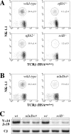
Figure 1.
Figure 1.
Reduced numbers of thymic NKT cells in NF-κB–deficient mice. Thymocytes from adult wild-type, nfkb1 / , nfkb2 / , and relB / (A) as well as from control and mIκBαtg mice (B) were analyzed for NK-1.1 and TCRβ expression by flow cytometry. Percentages of NKT cells (circles) were increased by gating on HSAneg-low cells. Numbers indicate mean values ± SD from 5–7 mice. (C) RT-PCR analysis of Vα14-Jα18 expression in wild-type, mIκBαtg, and relB / mice. Expression of the Cβ chain is shown as an amplification control.
Figure 2.
Figure 2.
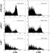
Selective loss of peripheral CD4+ NKT cells in relB / and nfkb1 / mice. NKT cells from adult wild-type, nfkb1 / , and relB / spleens were analyzed for CD4 and CD8 expression by flow cytometry. Histograms show CD4 (left) and CD8 (right) expression levels on NK-1.1+TCRβ+ cells. Note the reduction of CD4+ NKT cells in nfkb1 / and relB / mice and the increase of CD8+ NK-1.1+ T cells in nfkb1 / but not in relB / animals. Numbers indicate mean values ± SD from 3–7 mice.
Figure 3.
Figure 3.
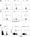
Analysis of NKT cell development in BM chimeras. (A) Thymocytes were isolated from lethally irradiated mice that were reconstituted with BM as indicated (donor BM → irradiated recipient). HSAneg-low cells were analyzed for NK-1.1 and TCRβ expression by flow cytometry. Percentages of NKT cells (mean values from 3–7 mice ± SD) are indicated. (B) Competitive BM chimeras were generated by mixing wild-type Ly-5.1 with mIκBαtg Ly-5.2 BM to reconstitute lethally irradiated wild-type Ly-5.2 mice. Chimerism was checked by flow cytometry (left). Thymocytes derived from wild-type (Ly-5.1+) and from mIκBαtg (Ly-5.2+) BM were stained for NK-1.1 and TCRβ expression and analyzed by flow cytometry (right). Percentages of NKT cells among total thymocytes (mean values from 2–3 mice ± SD) are indicated.
Figure 4.
Figure 4.
Analysis of IL-15 mRNA expression in thymus from NF-κB–deficient mice. RT-PCR analysis of whole thymus RNA revealed reduced IL-15 steady-state mRNA levels in relB / mice. Expression of IL-15 in thymus from mIκBαtg mice was similar to controls (unpublished data). Expression of the Cβ chain is shown as an amplification control.
Figure 5.
Figure 5.
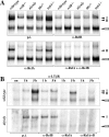
LTβR and NIK regulate RelB DNA-binding in thymus. (A) Whole thymus extracts from wild-type, relB / , aly/aly, ltbr / , and tnfr1 / mice were prepared and analyzed for κB-binding activity in EMSAs. Complexes are indicated by arrowheads and their identity was determined with Abs specific for individual Rel/NF-κB family members. p.i., preimmune serum; complex I, RelA heterodimers; complex II, RelB heterodimers. Complex III consists of p50 homodimers (unpublished data). (B) LTβR-mediated induction of RelB is dependent on NIK. Primary MEFs from wild-type and aly/aly mice were either left untreated (un) or treated for 1 and 5 h with anti-LTβR mAb and nuclear extracts were analyzed for κB-binding. Complexes were identified with specific Abs.
Figure 6.
Figure 6.
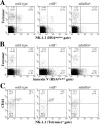
Analysis of developmental intermediates of Vα14i NKT cells in NF-κB-deficient mice. (A) Staining of thymocytes from 2-wk-old C57BL/6 wild-type, relB / , and mIκBαtg mice with anti-NK-1.1 and α-GalCer CD1d tetramer. (B) Analysis of apoptotic cells in thymus from 2-wk-old C57BL/6 wild-type, relB / , and mIκBαtg mice. Thymocytes were stained with Annexin V and α-GalCer CD1d tetramer. HSAneg-low cells in panels A and B were gated as in Fig. 1. (C) Expression of CD44 and NK-1.1 on tetramer+ thymocytes from 2-wk-old wild-type, relB / , and mIκBαtg mice was determined by flow cytometric analysis. Numbers in quadrants indicate mean values of percentages ± SD from three mice. (D) Numbers of tetramer+ cells in 106 thymocytes from 2-wk-old wild-type, relB / , and mIκBαtg mice based on their CD44 and NK-1.1 expression patterns.
Figure 6.
Figure 6.
Analysis of developmental intermediates of Vα14i NKT cells in NF-κB-deficient mice. (A) Staining of thymocytes from 2-wk-old C57BL/6 wild-type, relB / , and mIκBαtg mice with anti-NK-1.1 and α-GalCer CD1d tetramer. (B) Analysis of apoptotic cells in thymus from 2-wk-old C57BL/6 wild-type, relB / , and mIκBαtg mice. Thymocytes were stained with Annexin V and α-GalCer CD1d tetramer. HSAneg-low cells in panels A and B were gated as in Fig. 1. (C) Expression of CD44 and NK-1.1 on tetramer+ thymocytes from 2-wk-old wild-type, relB / , and mIκBαtg mice was determined by flow cytometric analysis. Numbers in quadrants indicate mean values of percentages ± SD from three mice. (D) Numbers of tetramer+ cells in 106 thymocytes from 2-wk-old wild-type, relB / , and mIκBαtg mice based on their CD44 and NK-1.1 expression patterns.
Figure 7.
Figure 7.
Model of NF-κB-regulated Vα14i NKT cell development in thymus. Lack of RelB in stromal cells results in impaired generation of CD44loNK-1.1 precursors. Blocking NF-κB in precursors results in impaired expansion of NKT cells predominantly at the NK-1.1 to NK-1.1+ transition. For more details see Discussion.

References

    1. Bendelac, A., M.N. Rivera, S.H. Park, and J.H. Roark. 1997. Mouse CD1-specific NK1 T cells: development, specificity, and function. Annu. Rev. Immunol. 15:535–562. - V体育平台登录 - PubMed
    1. Godfrey, D.I., K.J. Hammond, L.D. Poulton, M.J. Smyth, and A.G. Baxter. 2000. NKT cells: facts, functions and fallacies. Immunol. Today. 21:573–583. - V体育ios版 - PubMed
    1. MacDonald, H.R. 2002. Development and selection of NKT cells. Curr. Opin. Immunol. 14:250–254. - V体育官网入口 - PubMed
    1. Lantz, O., and A. Bendelac. 1994. An invariant T cell receptor α chain is used by a unique subset of major histocompatibility complex class I–specific CD4+ and CD4−8− T cells in mice and humans. J. Exp. Med. 180:1097–1106. - PMC - PubMed
    1. Bendelac, A., O. Lantz, M.E. Quimby, J.W. Yewdell, J.R. Bennink, and R.R. Brutkiewicz. 1995. CD1 recognition by mouse NK1+ T lymphocytes. Science. 268:863–865. - PubMed

Publication types

"VSports最新版本" MeSH terms