VSports手机版 - Skip to main page content
U.S. flag

An official website of the United States government

Dot gov

The VSports app下载. gov means it’s official. Federal government websites often end in . gov or . mil. Before sharing sensitive information, make sure you’re on a federal government site. .

Https

The site is secure. The https:// ensures that you are connecting to the official website and that any information you provide is encrypted and transmitted securely V体育官网. .

. 2003 May 1;22(9):2036-46.
doi: 10.1093/emboj/cdg189.

Ras promotes p21(Waf1/Cip1) protein stability via a cyclin D1-imposed block in proteasome-mediated degradation

Affiliations

VSports手机版 - Ras promotes p21(Waf1/Cip1) protein stability via a cyclin D1-imposed block in proteasome-mediated degradation

Mathew L Coleman et al. EMBO J. .

Abstract

Ras promotes the accumulation of the cyclin-dependent kinase inhibitor p21(Waf1/Cip1) (p21) VSports手机版. Previous studies reported that acute Raf/MEK/ERK activation elevates p21 protein levels by increased transcription. However, we have found that p21 induction in Ras-transformed murine fibroblasts occurs principally by a post-translational mechanism. Chronic activation of the Raf/MEK/ERK pathway blocked proteasome-mediated p21 degradation, resulting in accumulation of p21 protein with an elevated half-life. The stabilization of p21 by Ras was accompanied by high levels of p21-associated cyclin D1 and, similarly to Ras, cyclin D1 was sufficient to inhibit the proteasome-mediated p21 degradation. Knock-down of cyclin D1 by RNA interference confirmed that Ras-induced p21 stabilization was dependent upon cyclin D1 expression. We show that p21 directly binds to the C8alpha subunit of the 20S proteasome complex and that by competing for binding, cyclin D1 inhibits p21 degradation by purified 20S complexes in vitro. Therefore, we propose that Ras stabilizes p21 by promoting the formation of p21-cyclin D1 complexes that prevent p21 association with, and subsequent degradation by, the 20S proteasome. .


Figures
None
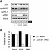
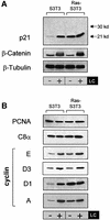
Fig. 1. Ras-induced p21 levels are uncoupled from the effects of proteasome inhibition and are elevated by a post-transcriptional mechanism. (A) Elevated p21 levels in Ras-transformed fibroblasts are not affected by proteasome inhibition. Parental (S3T3) and Ras-transformed S3T3 (Ras-S3T3) cells were serum starved for 16 h in the presence or absence of 10 µM lactacystin (LC) proteasome inhibitor. Extracts were western blotted as indicated. β-tubulin controlled for loading. (B) A subset of p21-binding partners are regulated by Ras in a manner similar to p21. Membranes shown in (A) were stripped and reprobed as indicated. (C) p21 mRNA is not elevated in Ras-S3T3 cells. Total RNA was isolated from serum-starved parental and Ras-S3T3 cells before northern blot analysis for p21 and cyclin D1. Expression levels were normalized to GAPDH levels and are presented as levels relative to the parental cell line.
None
Fig. 2. The Raf/MAPK effector pathway mediates Ras-induced post-transcriptional p21 regulation. (A) Inhibition of MEK reduces Ras-induced p21 levels and restores sensitivity to LC. Ras-S3T3 cells were incubated with 10 µM MEK inhibitor UO126 or 0.1% DMSO vehicle control and LC for 16 h. Extracts were western blotted as indicated. ERK2 controlled for loading. (B) p21 mRNA levels in Ras-S3T3 cells are not affected by UO126. Total RNA was isolated from Ras-S3T3 cells treated with UO126 or DMSO vehicle control for northern blot analysis of p21 and cyclin D1. Expression levels were normalized to GAPDH and are presented as levels relative to the parental cell line. (C) Raf activation is sufficient to uncouple p21 expression from the effects of proteasome inhibition. Raf:ER S3T3 cells treated with 100 nM 4-HT for 24–48 h were incubated with UO126 and/or LC in the absence of serum for 16 h. Extracts were western blotted as indicated. (D) Acute Raf activation elevates p21 and cyclin D1 mRNA. Total RNA was isolated from Raf:ER S3T3 cells treated as indicated and northern blotted for p21, cyclin D1 and GAPDH.
None
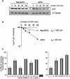
Fig. 3. Analyses of p21 protein stability. (A) p21 turnover is reduced in Ras-S3T3 versus parental S3T3 cells. S3T3 cells were treated with 25 µg/ml CHX to inhibit new protein synthesis for the times indicated, and extracts probed for p21 and β-tubulin to control for loading. A longer p21 exposure is shown for parental S3T3 cells to allow comparison. (B) p21 expression quantified by densitometric analysis. Expression is represented as the percentage remaining relative to time zero. Half-life values were calculated using lines of best fit. (C) Co-expression of Ras or cyclin D1 is sufficient to stabilize p21. Pulse–chase analysis was undertaken in NIH-3T3 cells transfected with epitope-tagged p21, H-Ras D12 and/or cyclin D1. Cells were treated with 10 µM UO126 (UO), 20 µM LY294002 (LY), 10 µM lactacystin (LC) or vehicle controls where indicated. Shown is the percentage of 35S-labelled p21 remaining after 2 h of chase, relative to a control sample lysed at time zero.
None
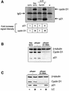
Fig. 4. Ras-induced cyclin D1 binds and stabilizes p21. (A) Ras and Raf signalling increase the proportion of p21 associated with cyclin D1. p21 was precipitated from S3T3. RAS-S3T3 or Raf:ER cells treated with or without 4-HT for 24 hours. As a control, Ras-S3T3 lysates were incubated with the M2 anti-flag antibody (*). Immunoprecipitated proteins were quantified using the relevant primary antibodies and a Cy5-coupled secondary IgG as described in Materials and methods. Shown below are the amounts of co-purified p21 and cyclin D1 relative to the control cell line (average of three experiments). (B) Cyclin D1 knock-down in Ras-S3T3 cells results in decreased p21. Lysates from parental Ras-S3T3 cells, two independent pSuper control clones and two independent cyclin D1 knock-down clones (pSuper D1 RNAi) were western blotted for β-tubulin, cyclin D1 and p21. (C) Knock-down of cyclin D1 restores p21 sensitivity to LC. One empty vector and one cyclin D1 knock-down clone were serum starved with or without LC treatment for 16 h as indicated, and lysates were blotted for cyclin D1 or p21.
None
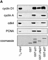
Fig. 5. Cyclin D1 binds to the p21 C-terminus and blocks the p21–C8α interaction. (A) Cyclin D1, but not cyclin A, binds to the Cy2 site of GST–p21. Ras-S3T3 extracts were incubated with the GST fusion proteins indicated, and captured complexes were western blotted for the p21-binding partners shown. A p21 mutant lacking the N-terminus was used (GST–ΔNp21) to determine which cyclins bind the C-terminal Cy2 site. (B) Association of p21 with C8α in vitro. IVTT-synthesized 35S-labelled p21 or luciferase control (L) were added to cyclin D1, C8α or Ral-His fusion proteins. Complexes were separated by SDS–PAGE, and His fusion-associated 35S-labelled proteins were detected by exposing membranes to autoradiography film. (C) Cyclin D1 competes with C8α for p21 binding. The assay described in (B) was repeated in the presence of 0, 2, 5 or 10 µg of cyclin D1, K-cyclin or GST–PCD fusions. Equal amounts of the different GST fusion proteins were added, as judged by western blot of one-tenth of the input.
None
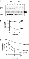
Fig. 6. Cyclin D1 inhibits 20S proteasome-mediated p21 degradation in vitro. (A) 20S proteasome activity degrades p21, but not p27, in vitro. IVTT p21 or p27 were incubated with 20S proteasome for the times indicated. Reactions were stopped with Laemmli buffer and analysed for 35S-labelled p21/p27 as described. (B) Proteasome-mediated degradation was quantified by PhosphorImager analysis of 35S-labelled proteins. Shown is the percentage of 35S-labelled protein remaining at each time point, relative to a sample at time zero. (C) Proteasome-mediated p21 degradation is blocked specifically by cyclin D1. The p21 degradation assay described in (A) was repeated in the presence of GST–cyclin D1 or GST–K-cyclin.
None
Fig. 7. Ras-induced stabilization of p21. (A) Low ERK activity. Under basal conditions, Ras-mediated cyclin D1 transcription and expression are low. The proportion of p21 bound to cyclin D1 is low and the C-terminus is free to bind the C8α subunit of the 20S proteasome. Constitutive proteasome-mediated degradation of p21 results in low levels of expression. (B) High ERK activity. Oncogenic mutation of Ras and constitutive activation of the Raf/ERK pathway drives high levels of cyclin D1 (or cyclin D1–cdk complexes) that may bind the C-terminal Cy2 site of p21, thereby masking the proteasome-binding motif. p21 association with C8α is blocked, thereby reducing 20S proteasome-mediated degradation.

References (V体育平台登录)

    1. Adams P.D., Sellers,W.R., Sharma,S.K., Wu,A.D., Nalin,C.M. and Kaelin,W.G.,Jr (1996) Identification of a cyclin–cdk2 recognition motif present in substrates and p21-like cyclin-dependent kinase inhibitors. Mol. Cell. Biol., 16, 6623–6633. - V体育安卓版 - PMC - PubMed
    1. Adnane J., Bizouarn,F.A., Qian,Y., Hamilton,A.D. and Sebti,S.M. (1998) p21WAF1/CIP1 is upregulated by the geranylgeranyltransferase I inhibitor GGTI-298 through a transforming growth factor β- and Sp1-responsive element: involvement of the small GTPase rhoA. Mol. Cell. Biol., 18, 6962–6970. - PMC (VSports最新版本) - PubMed
    1. Alessandrini A., Chiaur,D.S. and Pagano,M. (1997) Regulation of the cyclin-dependent kinase inhibitor p27 by degradation and phosphorylation. Leukemia, 11, 342–345. - PubMed
    1. Alt J.R., Gladden,A.B. and Diehl,J.A. (2002) p21Cip1 promotes cyclin D1 nuclear accumulation via direct inhibition of nuclear export. J. Biol. Chem., 277, 8517–8523. - PubMed
    1. Ball K.L., Lain,S., Fahraeus,R., Smythe,C. and Lane,D.P. (1997) Cell-cycle arrest and inhibition of Cdk4 activity by small peptides based on the carboxy-terminal domain of p21WAF1. Curr. Biol., 7, 71–80. - PubMed

Publication types

MeSH terms