Skip to main page content
U.S. flag

An official website of the United States government

Dot gov

The VSports app下载. gov means it’s official. Federal government websites often end in . gov or . mil. Before sharing sensitive information, make sure you’re on a federal government site. .

Https

The site is secure. The https:// ensures that you are connecting to the official website and that any information you provide is encrypted and transmitted securely V体育官网. .

. 2003 Apr 15;100(8):4389-94.
doi: 10.1073/pnas.0430973100. Epub 2003 Apr 3.

VSports手机版 - Domain-selective small-molecule inhibitor of histone deacetylase 6 (HDAC6)-mediated tubulin deacetylation

Affiliations

Domain-selective small-molecule inhibitor of histone deacetylase 6 (HDAC6)-mediated tubulin deacetylation

Stephen J Haggarty et al. Proc Natl Acad Sci U S A. .

Abstract

Protein acetylation, especially histone acetylation, is the subject of both research and clinical investigation. At least four small-molecule histone deacetylase inhibitors are currently in clinical trials for the treatment of cancer. These and other inhibitors also affect microtubule acetylation VSports手机版. A multidimensional, chemical genetic screen of 7,392 small molecules was used to discover "tubacin," which inhibits alpha-tubulin deacetylation in mammalian cells. Tubacin does not affect the level of histone acetylation, gene-expression patterns, or cell-cycle progression. We provide evidence that class II histone deacetylase 6 (HDAC6) is the intracellular target of tubacin. Only one of the two catalytic domains of HDAC6 possesses tubulin deacetylase activity, and only this domain is bound by tubacin. Tubacin treatment did not affect the stability of microtubules but did decrease cell motility. HDAC6 overexpression disrupted the localization of p58, a protein that mediates binding of Golgi elements to microtubules. Our results highlight the role of alpha-tubulin acetylation in mediating the localization of microtubule-associated proteins. They also suggest that small molecules that selectively inhibit HDAC6-mediated alpha-tubulin deacetylation, a first example of which is tubacin, might have therapeutic applications as antimetastatic and antiangiogenic agents. .


"V体育2025版" Figures

Figure 1
Figure 1
Characterization of tubacin, an inhibitor of α-tubulin deacetylation. (A) Summary of multidimensional screen of 7,392 small molecules having 1,3-dioxane diversity and deacetylase-biasing elements (14, 15). An image from wells showing selectivity of tubacin (yellow) in cytoblot assays measuring acetylated α-tubulin (AcTubulin) and acetylated lysine (AcLysine) is shown. (B) Chemical structure of tubacin (1) and an inactive analog niltubacin (2). An estimation of the logarithm of the partition coefficient between octanol and water (cLogP) predicts that both compounds have similar solubility properties. (C) Increased α-tubulin acetylation induced by tubacin in A549 cells detected by immunofluorescence with an antiacetylated α-tubulin antibody (red) and nuclear staining with Hoechst 33342 (blue). (D) Image from cytoblot assays detecting acetylated α-tubulin and acetylated lysine levels in A549 cells treated for 20 h with tubacin. (E) Selectivity of tubacin for inhibiting the TDAC versus HDACs determined by Western blot analysis of acetylated α-tubulin and histone H3 (K9 and K14) in A549 cells (5 h). The control compound niltubacin (2) showed no effect on α-tubulin or histone acetylation levels. A nonspecific band reactive with the acetylated histone H3 antibody was the loading control. (F) Quantification of the mean (n = 6) acetylated α-tubulin level in A549 cells after tubacin treatment using a cytoblot (estimated EC50 of 2.5 μM).
Figure 2
Figure 2
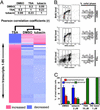
No effect of tubacin on gene expression or cell-cycle progression. (A) Pearson correlation matrix and nearest-neighbor clustering of a subset (86 of the most TSA-sensitive genes) of the average (n = 2) gene-expression data obtained from transcriptional profiling (murine U74Av2 gene chip; DCHIP software) of mouse embryonic stem cells treated (2.5 h) with TSA (300 nM) and tubacin (2 μM). (B) No effect of tubacin on DNA synthesis or cell-cycle distribution as determined by FACS analysis of BrdUrd-labeled and propidium iodide-stained A549 cells after treatment (4.5 h) with TSA (500 nM) and tubacin (2 μM). (C) Mitotic abnormalities in A549 cells treated (24 h) with HDAC inhibitors but not tubacin detected by immunofluorescence. The data are the average (±1 SD) of two treatments (≈100 mitotic cells per treatment).
Figure 3
Figure 3
Inhibition of HDAC6 deacetylase activity by tubacin. (A) Western blot of acetylated α-tubulin (AcTub) and total α-tubulin (α-Tub) after incubation of MAP-stabilized tubulin with wild-type HDAC6F and wild-type HDAC1F immunoprecipitated from transfected TAg Jurkat cells. Although HDAC1 showed no TDAC activity, the TDAC activity of HDAC6 was inhibited by TSA (300 nM) or tubacin (2 μM). (B) Decreased α-tubulin acetylation after tubacin (2 μM) treatment (4 h) of HDAC6-overexpressing compared with control (Neo) NIH 3T3 cells detected by immunofluorescence using antiacetylated α-tubulin (red) and anti-HDAC6 (green) antibodies and nuclear staining with Hoechst 33342 (blue). (C) Western blot of acetylated α-tubulin and total α-tubulin after incubation of MAP-stabilized tubulin with immunoprecipitants from transfected TAg Jurkat cells. Mutation of histidine 611 to alanine (H611A) but not histidine 216 to alanine (H216A) was sufficient to abolish the TDAC activity of HDAC6. (D) Effect of TSA and tubacin on the HDAC activity of immunoprecipitants from transfected TAg Jurkat cells. Activities (n = 3; ± mean absolute deviation) were measured by scintillation counting of 3H-labeled acetic acid released from 3H-labeled histones. Wild-type HDAC6F and the HDAC6F H216A mutant were inhibited only partially by TSA or tubacin, and the HDAC6F H611A mutant was not inhibited by either TSA or tubacin. (E) HDAC6 domain organization.
Figure 4
Figure 4
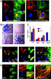
Phenotypic effects of tubacin. (A) No effect of tubacin (2 μM) pretreatment (4 h) on the stability of microtubules to nocodazole (332 nM; 2 h)-induced depolymerization determined by immunofluorescence using antiacetylated α-tubulin (red) and anti-α-tubulin (green) antibodies and nuclear staining with Hoechst 33342 (blue) in A549 cells. (B) Hematoxylin staining of NIH 3T3 cells (blue) that have undergone migration in a Transwell assay under the conditions indicated. Tubacin (2–20 μM) but not niltubacin (20 μM) inhibited cell migration. (C) Quantification of the migration of control (Neo) and HDAC6-overexpressing NIH 3T3 cells measured by counting hematoxylin-stained cells. Data are presented as the mean (n = 3; ±1 SD) relative to untreated wild-type (Neo) cells. ND, not determined. (D) Overexpression of HDAC6 but not catalytically inactive HDAC6 in NIH 3T3 cells causes mislocalization of p58 (arrowhead), a Golgi membrane MAP detected by immunofluorescence using anti-p58 (red) and anti-HDAC6 (green) antibodies and nuclear staining with Hoechst 33342 (blue). (E) Increased colocalization of HDAC6 with acetylated α-tubulin in NIH 3T3 cells (Neo) after tubacin (20 μM) treatment (4 h) detected by immunofluorescence using antiacetylated α-tubulin (red) and anti-HDAC6 (green) antibodies and nuclear staining with Hoechst 33342 (blue). The arrowhead indicates a region of higher magnification.

References

    1. Grozinger C M, Schreiber S L. Chem Biol. 2002;9:3–16. - PubMed
    1. Khochbin S, Verdel A, Lemercier C, Seigneurin-Berny D. Curr Opin Genet Dev. 2001;11:162–166. - PubMed (VSports手机版)
    1. Yoshida M, Kijima M, Akita M, Beppu T. J Biol Chem. 1990;265:17174–17179. - PubMed
    1. Kijima M, Yoshida M, Sugita K, Horinouchi S, Beppu T. J Biol Chem. 1993;268:22429–22435. - PubMed (VSports)
    1. Taunton J, Hassig C A, Schreiber S L. Science. 1996;272:408–411. - PubMed

Publication types

MeSH terms

LinkOut - more resources