Skip to main page content (V体育平台登录)
U.S. flag

An official website of the United States government

Dot gov

The . gov means it’s official. Federal government websites often end in . gov or . mil VSports app下载. Before sharing sensitive information, make sure you’re on a federal government site. .

Https

The site is secure. The https:// ensures that you are connecting to the official website and that any information you provide is encrypted and transmitted securely V体育官网. .

. 2003 Apr;71(4):1774-83.
doi: 10.1128/IAI.71.4.1774-1783.2003.

V体育官网入口 - Afa/Dr diffusely adhering Escherichia coli infection in T84 cell monolayers induces increased neutrophil transepithelial migration, which in turn promotes cytokine-dependent upregulation of decay-accelerating factor (CD55), the receptor for Afa/Dr adhesins

Affiliations

Afa/Dr diffusely adhering Escherichia coli infection in T84 cell monolayers induces increased neutrophil transepithelial migration, which in turn promotes cytokine-dependent upregulation of decay-accelerating factor (CD55), the receptor for Afa/Dr adhesins

V体育官网入口 - Fréderic Bétis et al. Infect Immun. 2003 Apr.

Abstract

Ulcerative colitis and Crohn's disease are inflammatory bowel diseases thought to involve strains of Escherichia coli VSports手机版. We report here that two wild-type Afa/Dr diffusely adhering E. coli (DAEC) strains, C1845 and IH11128, which harbor the fimbrial F1845 adhesin and the Dr hemagglutinin, respectively, and the E. coli laboratory strain HB101, transformed with the pSSS1 plasmid to produce Afa/Dr F1845 adhesin, all induced interleukin-8 (IL-8) production and transepithelial migration of polymorphonuclear leukocytes (PMNL) in polarized monolayers of the human intestinal cell line T84 grown on semipermeable filters. We observed that after PMNL migration, expression of decay-accelerating factor (DAF, or CD55), the brush border-associated receptor for Afa/Dr adhesins, was strongly enhanced, increasing the adhesion of Afa/Dr DAEC bacteria. When examining the mechanism by which DAF expression was enhanced, we observed that the PMNL transepithelial migration induced epithelial synthesis of tumor necrosis factor alpha and IL-1beta, which in turn promoted the upregulation of DAF. .


V体育官网 - Figures
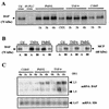
FIG. 1.
FIG. 1.
DAF is upregulated in T84 cells after f-MLP-induced PMNL transepithelial migration. (A) Western blot analysis revealed that the expression of DAF (CD55) in T84 cells after f-MLP-induced PMNL transmigration was greater than that in control T84 monolayers (Ctl). This upregulation was detected after 1 h of migration and peaked after 6 h of migration. The increased expression at 6 h of PMNL transmigration was not observed if the monolayers had been preincubated with cycloheximide (CHX). TNF-α treatment induced a marked increase in DAF expression after incubation of epithelial cells for 1 to 6 h. DAF upregulation was not observed in T84 monolayers infected for 1 or 3 h with strain C1845. DAF was not observed on T84 cells exposed to PI-PLC, which breaks down the GPI anchor of DAF. (B) Biotinylation assays showed that DAF was expressed only at the apical side (lanes A), not at the basolateral side (lanes Bl); either PMNL transepithelial migration for 2 h or exposure to TNF-α (2 h) induced increased DAF expression at the apical side of the T84 cells (relative to expression by control cells). Under these conditions, DAF expression also developed at the basolateral side of the T84 cells. MCP expression on T84 cells was used as a control: basolateral expression of MCP was not modified by TNF-α or PMNL transepithelial migration, and no MCP expression appeared at the apical side of the cells after PMNL transepithelial migration or TNF-α treatment (results of one of three experiments are shown). (C) DAF mRNA levels were increased in T84 cells after PMNL transmigration (3 or 6 h) or TNF-α treatment (3 or 6 h) but not in T84 monolayers infected for 1 or 3 h with strain C1845. In all panels, micrographs are representative of three to four experiments.
FIG. 2.
FIG. 2.
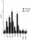
TNF-α and IL-1β production during the time-course of f-MLP-induced PMNL transepithelial migration (TM) in T84 cells. Cytokine production was measured by ELISAs. f-MLP-treated T84 cells or T84 cell monolayers infected for 1 or 3 h with strain C1845 served as negative controls. T84 cells treated with LPS or phorbol myristate acetate (PMA) served as positive controls (n = 4).
FIG. 3.
FIG. 3.
DAF upregulation observed in T84 monolayers during f-MLP-induced PMNL transmigration is linked to TNF-α and IL-1β production. DAF expression in T84 cells was analyzed by Western blotting after incubation of cells with or without MAbs against TNF-α and/or IL-1β added to the lower reservoir and subsequent PMNL transepithelial migration for 6 h. DAF upregulation observed after f-MLP-induced PMNL transepithelial migration or incubation of epithelial cells with TNF-α and/or IL-1β was decreased after treatment of T84 monolayers with either the anti-IL-1β or the anti-TNF-α MAb alone and was further decreased when the two MAbs were combined. In control (Ctl) cells, exposure to PI-PLC, which breaks down the GPI anchor of DAF, caused DAF to disappear. Micrographs are representative of three to four experiments.
FIG. 4.
FIG. 4.
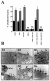
Quantification and observation by transmission electron microscopy of the attachment of Afa/Dr DAEC strains to T84 monolayers in the absence of or after PMNL transepithelial migration. (A) Cell-attached bacteria were distinguished from those that had entered the cells by treatment with gentamicin as described in Materials and Methods. T84 cell monolayers were incubated without or with wild-type Afa/Dr DAEC strain C1845 or IH11128 or with recombinant E. coli HB101-pSSS1 harboring Afa/Dr F1845 adhesin. E. coli K12-HB101 served as a negative control. The involvement of apical membrane-bound DAF in Afa/Dr adhesin attachment was investigated by using the anti-DAF MAb 1H4 and the isotype-matched control MAb MOPC21. Solid bars, bacterial attachment in control T84 monolayers after incubation with the bacteria for 2 h. Hatched bars, bacterial attachment after a 2-h incubation of T84 monolayers with the bacteria preceded by PMNL transmigration for 2 h. Data are expressed as the percentages of the inoculum that were associated with the cells. Data are means ± standard deviations from triplicate experiments. NS, not significantly different; ∗, P < 0.01. (B) Electron micrographs showing attachment of Afa/Dr DAEC bacteria to T84 monolayers in the absence of or after PMNL transepithelial migration. (B1) Control T84 monolayers; (B2) T84 monolayers incubated with IH11128 bacteria for 2 h; (B3) T84 monolayers incubated with IH11128 bacteria for 2 h followed by PMNL transepithelial migration for 2 h; (B4) T84 monolayers incubated with C1845 bacteria for 2 h followed by PMNL transepithelial migration for 2 h; (B5) C1845 bacteria adhering to T84 cells that had been exposed to MAb 1H4 before PMNL transepithelial migration for 2 h; (B6) C1845 bacteria adhering to T84 cells that had been exposed to MAb MOPC21 before PMNL transepithelial migration for 2 h. Arrows point to Afa/Dr DAEC bacteria adhering to the apical pole of the epithelial cells. Micrographs are representative of three experiments. Bars, 25 μm for panels B1 and B4; 10 μm for panels B2, B3, B5, and B6.
FIG. 5.
FIG. 5.
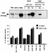
Adhesion of Afa/Dr DAEC strains to various epithelial cell lines is correlated with the level of DAF expression. (Top) Different levels of DAF expression in T84 (untreated or treated with EDTA), HeLa, HT-29, and CHO cell lines were analyzed by Western blotting. HT-29 cells, expressing DAF at a low level, and CHO cells, which did not express DAF, were first transfected with pcDNA-DAF and then subcultured at different times posttransfection for DAF expression. C, control; CHO Mt, CHO mutant without GPI expression. The immunoblot is representative of three experiments. (Bottom) Percentages of Afa/Dr DAEC strains (C1845 or IH11128 bacteria) adhering to the epithelial T84, HeLa, and HT29 cells after 6 h of incubation were strongly correlated with DAF levels. EDTA treatment of T84 monolayers before incubation with bacteria did not modify the percentage of adherent bacteria in comparison with that for control monolayers. In CHO cells, transfection of cDNA-DAF promoted Afa/Dr DAEC bacterial adhesion, a phenomenon abolished in CHO cells without GPI expression (CHO Mt). E. coli K12-HB101 was used as a control, and no binding was found in any of the cell lines used (data not shown). Data are expressed as the percentages of the inoculum that were associated with the cells. Data are means ± standard deviations from triplicate experiments.

References

    1. Abrami, L., M. Fivaz, T. Kobayashi, T. Kinoshita, R. G. Parton, and F. G. van der Goot. 2001. Cross-talk between caveolae and glycosylphosphatidylinositol-rich domains. J. Biol. Chem. 276:30729-30736. - "VSports注册入口" PubMed
    1. Andoh, A., Y. Fujiyama, K. Sumiyoshi, H. Sakumoto, and T. Bamba. 1996. Interleukin 4 acts as an inducer of decay-accelerating factor gene expression in human intestinal epithelial cells. Gastroenterology 111:911-918. - PubMed
    1. Andoh, A., Y. Fujiyama, K. Sumiyoshi, H. Sakumoto, H. Okabe, and T. Bamba. 1997. Tumour necrosis factor-alpha up-regulates decay-accelerating factor gene expression in human intestinal epithelial cells. Immunology 90:358-363. - PMC - PubMed
    1. Berin, M. C., A. Darfeuille-Michaud, L. J. Egan, Y. Miyamoto, and M. F. Kagnoff. 2002. Role of EHEC O157:H7 virulence factors in the activation of intestinal epithelial cell NF-κB and MAP kinase pathways and the upregulated expression of interleukin 8. Cell. Microbiol. 4:635-648. - PubMed
    1. Bernet-Camard, M. F., M. H. Coconnier, S. Hudault, and A. L. Servin. 1996. Differential expression of complement proteins and regulatory decay accelerating factor in relation to differentiation of cultured human colon adenocarcinoma cell lines. Gut 38:248-253. - "V体育ios版" PMC - PubMed

MeSH terms

LinkOut - more resources