Skip to main page content
U.S. flag

An official website of the United States government

Dot gov

The . gov means it’s official. Federal government websites often end in . gov or VSports app下载. mil. Before sharing sensitive information, make sure you’re on a federal government site. .

Https

The site is secure V体育官网. The https:// ensures that you are connecting to the official website and that any information you provide is encrypted and transmitted securely. .

. 2003 Mar;71(3):1513-9.
doi: 10.1128/IAI.71.3.1513-1519.2003.

"VSports app下载" Role of Toll-like receptor signaling in the apoptotic response of macrophages to Yersinia infection

Affiliations

Role of Toll-like receptor signaling in the apoptotic response of macrophages to Yersinia infection

Yue Zhang (VSports) et al. Infect Immun. 2003 Mar.

Abstract (V体育官网入口)

Macrophages encode several Toll-like receptors (TLRs) that recognize bacterial components, such as lipoproteins (TLR2) or lipopolysaccharides (TLR4), and activate multiple signaling pathways VSports手机版. Activation of transcription factor NF-kappaB by TLR2 or TLR4 signaling promotes proinflammatory and cell survival responses. Alternatively, TLR2 or TLR4 signaling can promote apoptosis if the activation of NF-kappaB is blocked. The gram-negative bacterial pathogen Yersinia pseudotuberculosis secretes into macrophages a protease (YopJ) that inhibits the activation of NF-kappaB and promotes apoptosis. We show that primary macrophages expressing constitutively active inhibitor kappaB kinase beta (IKKbeta) are completely resistant to YopJ-dependent apoptosis, indicating that YopJ inhibits signaling upstream of IKKbeta. Apoptosis is reduced two- to threefold in TLR4(-/-) macrophages infected with Y. pseudotuberculosis, while the apoptotic response of TLR2(-/-) macrophages to Y. pseudotuberculosis infection is equivalent to that of wild-type macrophages. Therefore, TLR4 is the primary source of apoptotic signaling in Yersinia-infected macrophages. Our results also show that a small percentage of macrophages can die as a result of an apoptotic process that is YopJ dependent but does not require TLR2 or TLR4 signaling. .


Figures

FIG. 1.
FIG. 1.
Strategy for the expression of wild-type or activated IKKβ in cells. (A) Structure of the bicistronic expression cassette for IKKβ and GFP in the retroviral expression vector. IKKβ contains an N-terminal FLAG epitope tag. The GFP control vector lacks sequences encoding IKKβ (not shown). (B) Kinase assay (KA) of IKKβ activity in transfected NIH 3T3 cells. NIH 3T3 cells transfected with retroviruses expressing wild-type IKKβ (IKKβ-wt) plus GFP, IKKβ-EE plus GFP, or GFP alone were left unstimulated or were stimulated with 25 ng of TNF-α/ml for 10 min after serum starvation. Detergent lysates of the cells were subjected to immunoprecipitation with anti-FLAG antibody. The immune complexes were incubated with purified GST- IκB(1- 62) in kinase assay buffer. Phosphorylation of GST- IκBα(1- 62) was monitored by Western blot analysis with phospho-specific IκBα antibody. The results shown are representative of three independent experiments.
FIG. 2.
FIG. 2.
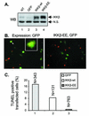
Constitutively active IKKβ protects macrophages from apoptosis induced by Yersinia infection. (A) Western blot (WB) analysis of IKKβ expression in macrophages. Nontransfected macrophages (NT) or macrophages transfected with retroviruses expressing wild-type IKKβ (IKKβ-wt) plus GFP, IKKβ-EE plus GFP, or GFP alone were lysed in detergent. Samples of the lysates were analyzed by Western blotting with anti-IKKα/β antibodies. Nonspecific bands (N.S.) indicate equal loading in the lanes. (B and C) TUNEL assay of Yersinia-induced apoptosis in transfected macrophages. Macrophages transfected with retroviruses expressing IKKβ-wt plus GFP, IKKβ-EE plus GFP, or GFP alone were infected with wild-type Y. pseudotuberculosis strain YP126 for 4 h. Apoptosis was detected by the TUNEL assay. Fluorescence microscopy was used to count the numbers of transfected macrophages (green cells) and the numbers of transfected macrophages that were TUNEL positive (red nuclei) in random nonoverlapping fields. In panel B, representative images of cells expressing GFP alone (left) or IKKβ-EE plus GFP (right) are shown. In the left image of panel B, two adjacent TUNEL-positive transfected macrophages are indicated by the arrowhead and enlarged in the inset. In panel C, the percentages of transfected macrophages that were scored as TUNEL positive are plotted. The data shown are from one representative experiment of three. N, number of transfected cells counted under each condition.
FIG. 3.
FIG. 3.
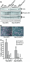
TLR4 contributes to Yersinia-induced signaling and apoptosis in macrophages. (A) Analysis of IκBα degradation and JNK phosphorylation in C3HeB/FeJ (WT) and C3H/HeJ (TLR4dn) macrophages. Macrophages were left untreated (basal), exposed to LPS (0.03, 0.1, 0.2, 0.3, or 10 μg/ml) or NaCl (0.2 M), or infected with YopJ Y. pseudotuberculosis (YP26) for 15 min. Cell lysates were subjected to Western blot analysis with anti-IκBα antibody (top), anti-phospho-JNK antibody (middle), or anti-JNK antibody (bottom). Two forms of JNK, p54 and p46, were detected. (B) Analysis of apoptosis by a TUNEL assay and fluorescence microscopy. Representative images show C3HeB/FeJ or C3H/HeJ macrophages stained by TUNEL after infection with wild-type (YP126) Y. pseudotuberculosis for 4 h. Red nuclei indicate a TUNEL-positive reaction. (C) Levels of YopJ-dependent apoptosis in C3HeB/FeJ or C3H/HeJ macrophages. The percentages of TUNEL-positive macrophages left uninfected or infected with wild-type (YP126) or YopJ (YP26) Y. pseudotuberculosis for 4 h are plotted. The results shown are representative of three independent experiments. N, number of cells counted under each condition.
FIG. 4.
FIG. 4.
TLR2, unlike TLR4, does not contribute to signaling or apoptosis in macrophages infected with Yersinia. (A) Analysis of IκBα degradation and JNK activation in wild-type (WT), TLR2−/−, or TLR4−/− bone marrow-derived macrophages. TLR2−/− mice were derived from 129/SvJ crossed with C57BL/6 (28), and TLR4−/− mice were derived from 129/Ola crossed with C57BL/6 (10, 28). Macrophages were left untreated (basal) or exposed to YopJ Y. pseudotuberculosis (YP26), LPS (0.1 or 0.3 μg/ml), or synthetic bacterial lipopeptide (BLP) (0.03 or 0.1 μg/ml) for 15 min. Cell lysates were subjected to Western blot analysis with anti-IκBα antibody (top), anti-phospho-JNK antibody (middle), or anti-JNK antibody (bottom). (B) Percentages of TUNEL-positive macrophages left uninfected (UI) or infected with wild-type (YP126) or YopJ (YP26) Y. pseudotuberculosis for 4 h. The results shown are representative of two experiments. N, number of cells counted.
FIG. 5.
FIG. 5.
Yersinia-induced signaling is delayed but not eliminated in TLR4−/− macrophages. Wild-type (WT), TLR2−/−, or TLR4−/− macrophages were infected with YopJ Y. pseudotuberculosis (YP26) for the indicated times in minutes. Cell lysates were prepared and subjected to Western blot (WB) analysis with anti-IκBα antibody (left), anti-phospho-JNK antibody (middle), or anti-JNK antibody (right). The results shown are representative of two experiments.

References

    1. Aderem, A., and R. J. Ulevitch. 2000. Toll-like receptors in the induction of the innate immune response. Nature 406:782-787. - V体育平台登录 - PubMed
    1. Akira, S., K. Takeda, and T. Kaisho. 2001. Toll-like receptors: critical proteins linking innate and acquired immunity. Nat. Immunol. 2:675-680. - "V体育官网入口" PubMed
    1. Aliprantis, A. O., R. B. Yang, D. S. Weiss, P. Godowski, and A. Zychlinsky. 2000. The apoptotic signaling pathway activated by Toll-like receptor-2. EMBO J. 19:3325-3336. - PMC - PubMed
    1. Celada, A., P. W. Gray, E. Rinderknecht, and R. D. Schreiber. 1984. Evidence for a gamma-interferon receptor that regulates macrophage tumoricidal activity. J. Exp. Med. 160:55-74. - PMC - PubMed
    1. Cornelis, G. R., A. Boland, A. P. Boyd, C. Geuijen, M. Iriarte, C. Neyt, M. P. Sory, and I. Stainier. 1998. The virulence plasmid of Yersinia, an antihost genome. Microbiol. Mol. Biol. Rev. 62:1315-1352. - PMC - PubMed

Publication types

MeSH terms

Substances

LinkOut - more resources