Skip to main page content
U.S. flag

An official website of the United States government

Dot gov

The . gov means it’s official. Federal government websites often end in VSports app下载. gov or . mil. Before sharing sensitive information, make sure you’re on a federal government site. .

Https

The site is secure. The https:// ensures that you are connecting to the official website and that any information you provide is encrypted and transmitted securely V体育官网. .

. 2002 Dec 24;99(26):16660-5.
doi: 10.1073/pnas.262589799. Epub 2002 Dec 16.

Reversal of DNA alkylation damage by two human dioxygenases

Affiliations

Reversal of DNA alkylation damage by two human dioxygenases (V体育ios版)

Tod Duncan et al. Proc Natl Acad Sci U S A. .

Abstract

The Escherichia coli AlkB protein protects against the cytotoxicity of methylating agents by repair of the DNA lesions 1-methyladenine and 3-methylcytosine, which are generated in single-stranded stretches of DNA. AlkB is an alpha-ketoglutarate- and Fe(II)-dependent dioxygenase that oxidizes the relevant methyl groups and releases them as formaldehyde. Here, we identify two human AlkB homologs, ABH2 and ABH3, by sequence and fold similarity, functional assays, and complementation of the E. coli alkB mutant phenotype. The levels of their mRNAs do not appear to correlate with cell proliferation but tissue distributions are different. Both enzymes remove 1-methyladenine and 3-methylcytosine from methylated polynucleotides in an alpha-ketoglutarate-dependent reaction, and act by direct damage reversal with the regeneration of the unsubstituted bases. AlkB, ABH2, and ABH3 can also repair 1-ethyladenine residues in DNA with the release of acetaldehyde. VSports手机版.


Figures (VSports最新版本)

Fig 1.
Fig 1.
Complementation of E. coli alkB mutant phenotype by overexpression of human ABH2 or ABH3 proteins, but not ABH1. Reactivation of MMS-treated φK single-stranded DNA phage was monitored in various E. coli strains. (A) Host strains were alkB+ or alkB22 derivatives of E. coli BL21.DE3. Plasmid pBAR54 encodes E. coli AlkB protein and pBAR65 encodes human ABH1 (not tagged). •, alkB+/pET15b; ◊, alkB22/pET15b; ▴, alkB22/pBAR54; □, alkB22/pBAR65. (B) Host strains were alkB+ or alkB22 derivatives of E. coli AB1157 gyrA. Plasmid pBAR32 encodes E. coli AlkB protein, pGST.ABH2 encodes GST tagged ABH2 and pGST.ABH3 encodes GST-tagged ABH3. •, alkB+/pGEX6P-1; ◊, alkB22/pGEX6P-1; ▴, alkB22/pBAR32; □, alkB22/pGST.ABH2; ▿, alkB22/pGST.ABH3. The proteins overexpressed in the alkB mutant are indicated with symbols in the corner of the figures.
Fig 2.
Fig 2.
(A) Approximate location of the core of the αKG–Fe(II) dioxygenase fold within AlkB, its putative human homologs, and three crystal structures from the αKG–Fe(II) dioxygenase superfamily: IPNS (isopenicillin N synthase from Aspergillus nidulans) (15), DAOCS (deacetoxycephalosporin C synthase from Streptomyces clavuligerus) (16), and ANS (anthocyanidin synthase from Arabidopsis thaliana) (17). (B) Multiple alignment within the core region of the AlkB homologs and the three crystal structures described above. Predicted secondary structures of ABH2 and ABH3 were calculated with the program PSI-PRED (19). The location of average secondary structure elements are shown as cylinders for α-helices and solid arrows for β-strands. These are colored green for known secondary structure locations (crystal structures) and red for predicted secondary structure locations (AlkB homologs). Residue background colors indicate the following properties: green, conserved hydrophobics; red, coordinate the Fe(II) ion; blue, conserved within the substrate binding pocket for all sequences; violet, conserved within the substrate binding pocket of the crystal structures only; magenta, conserved within the binding pocket for all AlkB homologs only; yellow, within the substrate binding pocket for all sequences but not conserved. (C) Three-dimensional model for the core region of ABH3. By using the known crystal structures of the three αKG-dependent DNA dioxygenases as templates and the alignment (B), a three-dimensional model was constructed for the core of ABH3 by using the program 3D-JIGSAW (24). The overall quality of side-chain packing and stereochemistry of the final model were checked by using program QUANTA 2000 (Accelrys, Orsay, France). The conserved α-helix is colored cyan. The β-strands, forming the jellyroll structure, are colored orange. The approximate location of the Fe(II) ion is represented as a white sphere. Side chains are colored according to the scheme described in B.
Fig 3.
Fig 3.
Repair of 1-methyladenine and 3-meC residues by ABH2 and ABH3. ABH2 or ABH3 (1 nmol) were incubated with [14C]MeI-treated poly(dC) (A and B) or poly(dA) (C and D) for 30 min under standard assay conditions. The 14C-labeled methylated bases remaining in the substrates were analyzed by HPLC and scintillation counting. ▵, no enzyme; □, ABH2 or ABH3.
Fig 4.
Fig 4.
Direct reversion of 3-meC to cytosine by E. coli AlkB and human ABH3. A thymine-rich oligodeoxynucleotide containing several [3H]cytosine residues was heavily methylated with DMS and then incubated with or without 0.23 nmol AlkB (A and B) or 0.84 nmol ABH3 (C and D) at 37°C for 30 min. Purified ABH3 was less active than AlkB; therefore, a reduced amount of substrate was incubated with ABH3 so that reversal of 3-meC to cytosine was readily seen. The [3H]cytosine residues remaining in the oligonucleotide were analyzed by HPLC and scintillation counting.
Fig 5.
Fig 5.
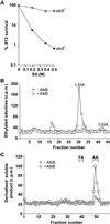
Repair of 1-ethyladenine by E. coli AlkB. (A) Defective reactivation of EtI treated M13 single-stranded DNA phage in an E. coli alkB mutant. Host strains were alkB+ and alkB117:Tn3 derivatives of F′/ABII57. ○, alkB+; •, alkB117:Tn3. (B) Removal of 1-etA from [14C]EtI-treated poly(dA) by 10 pmols AlkB protein. The 14C-ethylated adenines remaining in the substrate were examined by HPLC. ◊, No AlkB; □, +AlkB. (C) AlkB oxidizes 1-etA to release acetaldehyde. AlkB (10 pmols) was incubated with [14C]EtI-treated poly(dA), and the [14C]ethanol soluble material derivatized with DNPH. ◊, No AlkB; □, +AlkB. DNPH derivatives of formaldehyde and acetaldehyde were run as markers and monitored at A254.

References

    1. Bodell W. J. & Singer, B. (1979) Biochemistry 18, 2860-2863. - PubMed
    1. Boiteux S. & Laval, J. (1982) Biochimie 64, 637-641. - PubMed
    1. Kataoka H., Yamamoto, Y. & Sekiguchi, M. (1983) J. Bacteriol. 153, 1301-1307. - PMC - PubMed
    1. Sedgwick, B. & Lindahl, T. (2002) Oncogene Rev., in press. - "VSports在线直播" PubMed
    1. Chen B. J., Carroll, P. & Samson, L. (1994) J. Bacteriol. 176, 6255-6261. - PMC - PubMed

MeSH terms

LinkOut - more resources