Skip to main page content
U.S. flag

An official website of the United States government

Dot gov

The . gov means it’s official. Federal government websites often end in . gov or . mil. Before sharing sensitive information, make sure you’re on a federal government site VSports app下载. .

Https

The site is secure. The https:// ensures that you are connecting to the official website and that any information you provide is encrypted and transmitted securely. V体育官网.

. 2002 Sep 2;21(17):4621-31.
doi: 10.1093/emboj/cdf461.

TIS7 interacts with the mammalian SIN3 histone deacetylase complex in epithelial cells

Affiliations

TIS7 interacts with the mammalian SIN3 histone deacetylase complex in epithelial cells

Ilja Vietor et al. EMBO J. .

V体育安卓版 - Abstract

The mammalian SIN3 complex consists of histone deacetylases (HDAC1, HDAC2), several known proteins (SAP30, N-CoR) and as yet unidentified proteins. Here we show that the mouse tetradecanoyl phorbol acetate induced sequence 7 (TIS7) protein is a novel transcriptional co-repressor that can associate with the SIN3 complex VSports手机版. We have identified tis7 as a gene that is up-regulated upon loss of polarity in a mouse mammary gland epithelial cell line expressing an estrogen-inducible c-JunER fusion protein. In unpolarized cells, TIS7 protein levels increase and TIS7 translocates into the nucleus. Overexpression of tis7 causes loss of polarity and represses a set of genes, as revealed by cDNA microarray analysis. We have shown that TIS7 protein interacts with several proteins of the SIN3 complex (mSin3B, HDAC1, N-CoR and SAP30) by yeast two-hybrid screening and co-immunoprecipitations. TIS7 co-immunoprecipitated HDAC complex is enzymatically active and represses a GAL4-dependent reporter transcription. The transcriptional repression of endogenous genes by tis7 overexpression is HDAC dependent. Thus, we propose TIS7 as a transcriptional co-repressor affecting the expression of specific genes in a HDAC activity-dependent manner during cell fate decisions, e. g. scattering. .


Figures

None
Fig. 1. TIS7 mRNA and protein levels are up-regulated and TIS7 protein translocates into the nucleus of E2-activated c-JunER cells. (A) Northern blot analysis of total RNA isolated from c-JunER cells hybridized with a TIS7 cDNA probe. Control cells (–), 48 h E2-treated cells (+), reverted cells (Rev = cells grown for 48 h with E2 and then cultured for two additional passages in the absence of E2). Northern blot analysis of EpH4 parental cell line. One representative northern blot from five (c-JunER cells) or seven (EpH4 cells) independent experiments is shown. (B) Specificity of the affinity-purified TIS7 rabbit polyclonal antibodies. Left panel: GST–TIS7 fusion proteins detected by western blotting using α-GST antibodies. Middle panel: same proteins analysed using the affinity-purified α-TIS7 antibodies. Right panel: immunoprecipitation experiment using same the α-TIS7 antibodies without (–peptide) or after pre-incubation with the peptide used for immunization. HeLa cells were transfected with Xpr–TIS7 vector construct and equal amounts of cell lysate were used for immunoprecipitation. Immunoprecipitated protein Xpr–TIS7 was detected by western blotting using α-Xpress monoclonal antibodies. (C) Western blot analysis of TIS7 protein in control (–E2) and E2-treated (+E2) c-JunER cells, respectively. Equal loading is documented by the actin immunostaining (bottom). Western blot images were scanned, quantified and are presented as a ratio between the intensity of the specific TIS7 and actin input control signals (mean ± SD, n = 3). (D) Laser scanning confocal immunofluorescence analysis of TIS7 and ZO-1 localization in c-JunER polarized cells (–E2; left panel) and cells treated with 10–6 M E2 for 4 days (right panels). Loss of polarity of the epithelial cells was followed by measuring the transepithelial resistance (TER values). Size bar = 10 µm.
None
Fig. 2. TIS7-overexpressing cells lose epithelial polarity. (A) Cells were infected with CELO-recombinant viruses. Overexpressed TIS7 was detected by laser scanning confocal immunofluorescence microscopy using α-Myc antibody, endogenous β-catenin with the monoclonal antibody and eGFP directly. Top panels show sections through the xy planes and underneath the xz side view is shown. Bar = 10 µm. (B) TIS7-overexpressing cell grows on the top of the monolayer of otherwise polarized epithelial cells (arrowhead marks the filter insert). In polarized cells, tight junctional protein ZO-1 forms a typical ring-like structure (arrow). (C) TIS7 overexpression caused a significant decrease in the transepithelial resistance.
None
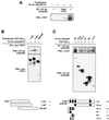
Fig. 3. TIS7 interacts with mSin3B. (A) HeLa cells were transfected with FLAG-mSin3B LF expression plasmid. Cell lysates were immunoprecipitated with α-FLAG tag monoclonal antibody and co-immunoprecipitated endogenous TIS7 was detected by western blot analysis. (B) HeLa cells were co-transfected with N- or C-terminal Myc9-tagged TIS7 truncation mutants and FLAG-tagged mSin3B long-form construct as indicated. Cell lysates were immunoprecipitated with the antibodies against the Myc tag, and co-immunoprecipitated mSin3B was detected by immunoblotting using α-FLAG antibodies. The bottom panel shows TIS7-Myc9 truncation mutants immunoprecipitated with the α-Myc antibodies. (C) HeLa cells were co-transfected with C-terminally Myc9-tagged full-length TIS7 construct and different N-terminally FLAG-tagged mSin3B constructs (LF, long form; P, PAH domain; arrowheads point towards the Sin3 fusion protein bands, smaller bands are degradation products). Protein complexes were immunoprecipitated with α-FLAG tag antibodies. In the lower panel are mSin3B constructs immunoprecipitated with α-FLAG antibodies detected with the same antibody. Schemes under both panels depict expressed parts of the proteins and level of interaction (–, +, ++).
None
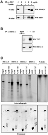
Fig. 4. TIS7 interacts with HDAC1. (A) HeLa cell lysates were immunoprecipitated with the affinity-purified rabbit α-TIS7 antibodies, without (–) or with (+) pre-incubation with the peptide used to raise the TIS7 antibodies (left panel). Co-immunoprecipitated HDAC1 was detected by western blotting. Increasing amounts of the α-TIS7 antibodies co-immunoprecipitated increasing amounts of the endogenous HDAC1. Bottom panels show immunoprecipitated endogenous TIS7 detected by western blotting using the same α-TIS7 antibodies. (B) In a vice versa experiment, HeLa cell lysates were immunoprecipitated with the monoclonal antibodies against HDAC1 and co-immunoprecipitated endogenous TIS7 was detected by western blotting (–Ab, pre-immune serum). The bottom panel documents amounts of immunoprecipitated endogenous HDAC1. (C) GST pull-down analysis using recombinant GST–TIS7 protein. 35S-labelled HDAC1, HDAC2, HDAC3, hSCC1 and N-CoR products of in vitro coupled transcription/translation reaction in rabbit reticulocyte lysate were incubated with GST proteins bound to the glutathione beads, washed and eluted with sample buffer. Bound proteins were analysed by autoradiography. Equal loading of GST and GST–TIS7 proteins was confirmed by Coomassie Blue staining (bottom panel).
None
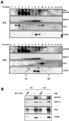
Fig. 5. TIS7 can associate with the SIN3 complex. (A) mSin3B, HDAC1, TIS7 and SAP30 partially co-fractionate. Nuclear extracts prepared from control (–E2) or 4 day E2-treated c-JunER cells (+E2) were fractionated on a 10–35% continuous sucrose gradient. Aliquots from 0.6 ml fractions were resolved on a 10% SDS–PAGE gel and immunoblotted with the indicated antibodies. In mSin3B western blot, the arrowhead indicates the full-length mSin3B LF band; other bands detected by this antibody, mainly in lighter fractions, represent degradation products. Sedimentation coefficients of 14 and 20S fractions were marked according to the peaks of proteasome and cohesin proteins used as markers. (B) c-JunER cells were either left untreated (–E2) or cultured in the presence of 10–6 M E2 for 4 days. Nuclear extracts were prepared as described in Dignam et al. (1983), and immunoprecipitated with the affinity-purified α-TIS7 antibody. Co-immunoprecipitated endogenous proteins were detected by western blotting with the indicated antibodies. The additional band in the TIS7 western blot is an as yet unidentified post-translational modification present in E2-treated cells.
None
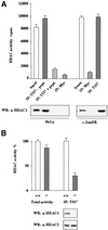
Fig. 6. TIS7 co-immunoprecipitated HDAC is enzymatically active. (A) HeLa and c-JunER cell lysates (as indicated below the graphs) were immunoprecipitated with affinity-purified α-TIS7 antibodies, without (–pept.) or with TIS7 N-terminal peptide pre-incubation (+ pept.), or with unrelated antibodies (α-Myc) as a negative control. HDAC activity was measured as described previously (Doetzlhofer et al., 1999). HDAC1 was detected by western blot analysis with mouse monoclonal α-HDAC1 antibodies. The input lane represents 4% of the total lysate used for the immunoprecipitation and documents the functionality of the enzymatic assay. This experiment was repeated several times with similar results; one representative experiment is shown, values of the bars are mean ± SD, n = 3. (B) TIS7 interacts mainly with HDAC1. The left side of the graph shows the HDAC enzymatic activity of total cell lysates derived from wild-type and homozygous HDAC1–/– embryonic stem cells. The right side of the graph shows the result of the HDAC enzymatic activity measured in the same cell lysates following the immunoprecipitation with affinity-purified α-TIS7 antibodies. The western blot underneath the graph shows HDAC1 and HDAC2 detected using monoclonal antibodies. (C) TIS7 inhibits transcriptional activity. HeLa cells were transiently co-transfected with the pLGCLuc-E351 luciferase reporter construct, β-galactosidase expression plasmid and either GAL4 DNA-binding domain (DBD) alone or GAL4-DBD-TIS7 wild-type, N- (1–321; 1–367) and C-terminal (140–449) TIS7 plasmid, respectively. After 36 h, the transfected cells were treated with 50 ng/ml TSA or left untreated, and 12 h later luciferase and β-galactosidase activities were measured. Luciferase intensities were normalized to β-galactosidase activity. Values of cells transfected with pPuroGAL4-DBD empty vector, reporter construct and not treated with TSA represented 100%.
None
Fig. 7. TIS7-repressed gene expression is HDAC dependent. c-JunER cells were infected with CELO-eGFP or CELO-TIS7. After 36 h, cells were treated with 10 ng/ml TSA (+TSA) or left untreated (–TSA). After an additional 12 h, total RNAs were isolated and expression of crabp II was measured by Real-Time PCR. Expression of crabp II was normalized to the expression of gapdh. Western blots at the bottom equal expression of eGFP or TIS7 protein in both samples.

References

    1. Ahringer J. (2000) NuRD and SIN3 histone deacetylase complexes in development. Trends Genet., 16, 351–356. - PubMed (V体育官网)
    1. Alkema M.J., Jacobs,J., Voncken,J.W., Jenkins,N.A., Copeland,N.G., Satijn,D.P., Otte,A.P., Berns,A. and van Lohuizen,M. (1997) MPc2, a new murine homolog of the Drosophila polycomb protein is a member of the mouse polycomb transcriptional repressor complex. J. Mol. Biol., 273, 993–1003. - PubMed
    1. Alland L., Muhle,R., Hou,H.,Jr, Potes,J., Chin,L., Schreiber-Agus,N. and DePinho,R.A. (1997) Role for N-CoR and histone deacetylase in Sin3-mediated transcriptional repression. Nature, 387, 49–55. - PubMed
    1. Ayer D.E. (1999) Histone deacetylases: transcriptional repression with SINers and NuRDs. Trends Cell Biol., 9, 193–198. - "V体育安卓版" PubMed
    1. Ayer D.E., Lawrence,Q.A. and Eisenman,R.N. (1995) Mad-Max transcriptional repression is mediated by ternary complex formation with mammalian homologs of yeast repressor Sin3. Cell, 80, 767–776. - PubMed

Publication types

MeSH terms

Substances

Associated data