Skip to main page content
U.S. flag

An official website of the United States government

Dot gov

The . gov means it’s official. Federal government websites often end in . gov or . mil. Before sharing sensitive information, make sure you’re on a federal government site. VSports app下载.

Https

The site is secure. The https:// ensures that you are connecting to the official website and that any information you provide is encrypted and transmitted securely. V体育官网.

. 2002 Aug 5;196(3):323-33.
doi: 10.1084/jem.20011612.

Uncoupling of proliferative potential and gain of effector function by CD8(+) T cells responding to self-antigens

Affiliations

Uncoupling of proliferative potential and gain of effector function by CD8(+) T cells responding to self-antigens

Javier Hernández et al. J Exp Med. .

Abstract

Professional antigen-presenting cells (APCs) are capable of transporting self-antigens from peripheral tissues to secondary lymphoid organs where they are presented to potentially autoreactive CD8(+) T cells. In the absence of an inflammatory response, this results in immune tolerance. The presence of activated, antigen-specific CD4(+) T cells converts this tolerogenic encounter into an immunogenic one by promoting extensive proliferation of CD8(+) T cells and their development into effectors. Surprisingly, activation of APCs with an agonistic antibody specific for CD40 could not substitute for CD4(+) help in this task. Anti-CD40 induced recruitment of dendritic cells expressing high levels of B7 costimulatory molecules into the lymph nodes, which in turn, greatly enhanced activation and expansion of CD8(+) T cells. However, these activated CD8(+) cells did not demonstrate effector function VSports手机版. We conclude that proliferative potential and gain of effector function are separable events in the differentiation program of CD8(+) T cells. .


Figures

Figure 1.
Figure 1.
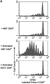
In vivo and in vitro proliferation of HNT CD4+ T cells. (A) 3 × 10⁶ CFSE-labeled, purified, Thy1.1+ clone 4 CD8+ T cells (top) or HNT CD4+ T cells (bottom) were injected into InsHA hosts. Mice were killed on day 4 after transfer and cells from LNs were analyzed by FACS®. Histograms represent the amount of CFSE label gating on CD8+ Thy1.1+ lymphocytes (top) or CD4+ lymphocytes (bottom) of one representative experiment of five independent ones for clone 4 and three for HNT. The unstained cells in the bottom panels represent the endogenous CD4+ T cell repertoire. (B) Purified, CFSE-labeled HNT or DO11 CD4+ T cells were activated in vitro for 3 d and analyzed by FACS®. Histograms represent the amount of CFSE label gating on live CD4+ T cells.
Figure 1.
Figure 1.
In vivo and in vitro proliferation of HNT CD4+ T cells. (A) 3 × 10⁶ CFSE-labeled, purified, Thy1.1+ clone 4 CD8+ T cells (top) or HNT CD4+ T cells (bottom) were injected into InsHA hosts. Mice were killed on day 4 after transfer and cells from LNs were analyzed by FACS®. Histograms represent the amount of CFSE label gating on CD8+ Thy1.1+ lymphocytes (top) or CD4+ lymphocytes (bottom) of one representative experiment of five independent ones for clone 4 and three for HNT. The unstained cells in the bottom panels represent the endogenous CD4+ T cell repertoire. (B) Purified, CFSE-labeled HNT or DO11 CD4+ T cells were activated in vitro for 3 d and analyzed by FACS®. Histograms represent the amount of CFSE label gating on live CD4+ T cells.
Figure 2.
Figure 2.
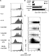
Activated HNT CD4+ T cells enhance proliferation and effector function of clone 4 CD8+ T cells in the pancreatic LNs of InsHA mice. 3 × 10⁶ CFSE-labeled, purified, Thy1.1+ clone 4 CD8+ T cells were injected into InsHA hosts either alone or along with nonlabeled, 3 × 10⁶ purified HNT CD4+ T cells, 1.5 × 10⁶ in vitro activated HNT CD4+ T cells or 1.5 × 10⁶ in vitro activated DO11 CD4+ T cells, as indicated. These data is representative of two to five independent experiments including a total of at least six mice per group per time point. (A) Mice were killed on day 4 after transfer and cells from pancreatic LNs were analyzed by FACS®. Histograms represent the amount of CFSE label gating on CD8+ Thy1.1+ lymphocytes. (B) Total numbers of CD8+ Thy1.1+ cells in the pancreatic LNs of host mice killed on days 4 and 8 after transfer. Data represent the mean of all experiments performed. Only negative standard deviation is depicted to achieve greater sensitivity in the graph. (C) On day 4 after transfer cells from pancreatic LNs were incubated with Kd HA peptide for 6 h and then analyzed by FACS® to detect accumulation of intracellular IFN-γ. Plots represent the amount of CFSE label versus the intensity of IFN-γ produced gating on lymphocytes CD8+ Thy1.1+. The percentage of clone 4 IFN-γ+ cells is indicated. The percentage of IFN-γ+ cells in controls that were stimulated with an irrelevant peptide was >1% in all the cases.
Figure 2.
Figure 2.
Activated HNT CD4+ T cells enhance proliferation and effector function of clone 4 CD8+ T cells in the pancreatic LNs of InsHA mice. 3 × 10⁶ CFSE-labeled, purified, Thy1.1+ clone 4 CD8+ T cells were injected into InsHA hosts either alone or along with nonlabeled, 3 × 10⁶ purified HNT CD4+ T cells, 1.5 × 10⁶ in vitro activated HNT CD4+ T cells or 1.5 × 10⁶ in vitro activated DO11 CD4+ T cells, as indicated. These data is representative of two to five independent experiments including a total of at least six mice per group per time point. (A) Mice were killed on day 4 after transfer and cells from pancreatic LNs were analyzed by FACS®. Histograms represent the amount of CFSE label gating on CD8+ Thy1.1+ lymphocytes. (B) Total numbers of CD8+ Thy1.1+ cells in the pancreatic LNs of host mice killed on days 4 and 8 after transfer. Data represent the mean of all experiments performed. Only negative standard deviation is depicted to achieve greater sensitivity in the graph. (C) On day 4 after transfer cells from pancreatic LNs were incubated with Kd HA peptide for 6 h and then analyzed by FACS® to detect accumulation of intracellular IFN-γ. Plots represent the amount of CFSE label versus the intensity of IFN-γ produced gating on lymphocytes CD8+ Thy1.1+. The percentage of clone 4 IFN-γ+ cells is indicated. The percentage of IFN-γ+ cells in controls that were stimulated with an irrelevant peptide was >1% in all the cases.
Figure 3.
Figure 3.
Anti-CD40 treatment enhances proliferation but not effector function of clone 4 CD8+ T cells in the pancreatic LNs of InsHA mice. 3 × 10⁶ CFSE-labeled, purified, Thy1.1+ clone 4 CD8+ T cells were injected into InsHA or BALB/c hosts. Mice were then treated with anti-CD40 mAb or purified rat IgG as isotype control. Anti-CD40 treated mice and isotype controls were then either left without further treatment or treated with IL-2 or IL-12 as described in Materials and Methods and as indicated in the figure. These data is representative of two to five independent experiments including a total of at least six mice per group per time point. (A) Proliferation profiles of clone 4 CD8+ T cells on day 4 after transfer were determined as in Fig. 2 A. (B) Total numbers of CD8+ Thy1.1+ cells in the pancreatic LNs of host mice killed on days 4 or 8 after transfer determined as in Fig. 2 B. (C) Production of IFN-γ on day 4 after transfer was determined as in Fig. 2 C.
Figure 4.
Figure 4.
Blockade of B7 interaction inhibits accumulation and effector function of clone 4 CD8 T cells. (A) 3 × 10⁶ CFSE-labeled, purified, Thy1.1+ clone 4 CD8+ T cells were injected into InsHA recipients along with anti-CD40 mAb or with 1.5 × 10⁶ in vitro activated HNT CD4+ T cells. Half of the mice from the previous groups were treated with anti-B7.1 plus anti-B7.2 mAbs and the rest were treated with a mixture of rat and hamster IgG as isotype controls. On day 4 after transfer total numbers of CD8+ Thy1.1+ cells in the pancreatic LNs of host mice were determined as in Fig. 2 B. Results represent data from six mice per group analyzed in two independent experiments. The line represent the number of clone 4 CD8+ T cells in mice that received clone 4 CD8+ T cells without anti-CD40 or HNT cells. (B) Production of IFN-γ on day 4 after transfer was determined as in Fig. 2 C.
Figure 4.
Figure 4.
Blockade of B7 interaction inhibits accumulation and effector function of clone 4 CD8 T cells. (A) 3 × 10⁶ CFSE-labeled, purified, Thy1.1+ clone 4 CD8+ T cells were injected into InsHA recipients along with anti-CD40 mAb or with 1.5 × 10⁶ in vitro activated HNT CD4+ T cells. Half of the mice from the previous groups were treated with anti-B7.1 plus anti-B7.2 mAbs and the rest were treated with a mixture of rat and hamster IgG as isotype controls. On day 4 after transfer total numbers of CD8+ Thy1.1+ cells in the pancreatic LNs of host mice were determined as in Fig. 2 B. Results represent data from six mice per group analyzed in two independent experiments. The line represent the number of clone 4 CD8+ T cells in mice that received clone 4 CD8+ T cells without anti-CD40 or HNT cells. (B) Production of IFN-γ on day 4 after transfer was determined as in Fig. 2 C.
Figure 5.
Figure 5.
Anti-CD40 treatment promotes enhanced migration of activated CD11c+ cells to the pancreatic LNs of InsHA mice. Groups of InsHA mice were treated with anti-CD40 or rat IgG isotype control polyclonal Ab. On day 4, cells from the pancreatic LN were analyzed by FACS® for the expression of CD11c and B7.1 or B7.2 molecules. (A) Total numbers of CD11c+ cells in the pancreatic LNs. Data represent the mean and standard deviation of seven mice analyzed in two independent experiments. (B) Histograms represent the expression of B7 molecules gating in CD11c+ cells from the pancreatic LNs of representative treated InsHA mice. The geometrical mean fluorescence intensity is indicated in each panel.
Figure 5.
Figure 5.
Anti-CD40 treatment promotes enhanced migration of activated CD11c+ cells to the pancreatic LNs of InsHA mice. Groups of InsHA mice were treated with anti-CD40 or rat IgG isotype control polyclonal Ab. On day 4, cells from the pancreatic LN were analyzed by FACS® for the expression of CD11c and B7.1 or B7.2 molecules. (A) Total numbers of CD11c+ cells in the pancreatic LNs. Data represent the mean and standard deviation of seven mice analyzed in two independent experiments. (B) Histograms represent the expression of B7 molecules gating in CD11c+ cells from the pancreatic LNs of representative treated InsHA mice. The geometrical mean fluorescence intensity is indicated in each panel.

References

    1. Stockinger, B. 1999. T lymphocyte tolerance: from thymic deletion to peripheral control mechanisms. Adv. Immunol. 71:229–265. - PubMed
    1. Heath, W.R., and F.R. Carbone. 2001. Cross-presentation, dendritic cells, tolerance and immunity. Annu. Rev. Immunol. 19:47–64. - V体育官网 - PubMed
    1. Albert, M.L., B. Sauter, and N. Bhardwaj. 1998. Dendritic cells acquire antigen from apoptotic cells and induce class I-restricted CTLs. Nature. 392:86–89. - PubMed
    1. Huang, F.P., N. Platt, M. Wykes, J.R. Major, T.J. Powell, C.D. Jenkins, and G.G. MacPherson. 2000. A discrete subpopulation of dendritic cells transports apoptotic intestinal epithelial cells to T cell areas of mesenteric lymph nodes. J. Exp. Med. 191:435–444. - "VSports app下载" PMC - PubMed
    1. Kurts, C., M. Cannarile, I. Klebba, and T. Brocker. 2001. Dendritic cells are sufficient to cross-present self-antigens to CD8 T cells in vivo. J. Immunol. 166:1439–1442. - PubMed

Publication types (VSports在线直播)

MeSH terms