Skip to main page content
U.S. flag

An official website of the United States government

Dot gov

The . gov means it’s official. Federal government websites often end in . gov or . mil. Before sharing sensitive information, make sure you’re on a federal government site. VSports app下载.

Https

The site is secure. The https:// ensures that you are connecting to the official website and that any information you provide is encrypted and transmitted securely. V体育官网.

. 2002 Jul;184(14):3898-908.
doi: 10.1128/JB.184.14.3898-3908.2002.

Identification of a new class of cytochrome P450 from a Rhodococcus sp (V体育官网入口)

Affiliations

Identification of a new class of cytochrome P450 from a Rhodococcus sp

Gareth A Roberts et al. J Bacteriol. 2002 Jul.

Abstract

A degenerate set of PCR primers were used to clone a gene encoding a cytochrome P450 (the P450RhF gene) from Rhodococcus sp. strain NCIMB 9784 which is of unique primary structural organization. Surprisingly, analysis of the translation product revealed that the P450 is fused to a reductase domain at the C terminus which displays sequence conservation for dioxygenase reductase proteins. The reductase partner comprises flavin mononucleotide- and NADH-binding motifs and a [2Fe2S] ferredoxin-like center. The gene was engineered for heterologous expression in Escherichia coli, and conditions were found in which the enzyme was produced in a soluble form. A recombinant strain of E. coli was able to mediate the O dealkylation of 7-ethoxycoumarin in good yield, despite the absence of any recombinant redox proteins. This unprecedented finding leads us to propose that P450RhF represents the first example of a new class of cytochromes P450 in which the reducing equivalents are supplied by a novel reductase in a fused arrangement. VSports手机版.


Figures

FIG. 1.
FIG. 1.
Schematic representation of the different classes of cytochrome P450 systems. Class I systems comprise a FAD-containing flavodoxin reductase, an iron-sulfur protein (ferredoxin), and the P450. The eukaryotic class I enzymes are associated with the mitochondrial membrane. In a class II system, the P450 is partnered with a diflavin reductase, whereas in the class III system, the diflavin reductase is fused to the P450. We propose that the new class IV system is made up of an FMN-containing reductase with a ferredoxin-like center linked to a P450 in a single polypeptide. The N and C termini of the fused class III and IV systems are shown.
FIG. 2.
FIG. 2.
Restriction map of a 4.4-kbp segment of chromosomal DNA from Rhodococcus sp. strain NCIMB 9784 containing the P450RhF gene. ORFs are denoted by arrows, which indicate the direction of transcription. The relative positions of the three overlapping clones are shown by the thick bars below the restriction map. P350 represents the PCR product obtained by using a degenerate set of primers designed from the oxygen- and heme-binding motifs of cytochrome P450 enzymes. The asterisks denote the site from which an oligonucleotide hybridization probe (P45) was designed. This probe was then used to clone both Sm3.0 and Bc3.5 from a sublibrary of genomic DNA. Only the restriction sites mentioned in the text are shown.
FIG. 3.
FIG. 3.
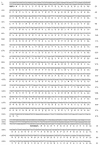
DNA sequence of the 4,411-bp SmaI-BclI region encoding the cytochrome P450 (P450RhF) from Rhodococcus sp. strain NCIMB 9784. The deduced ORFs are labeled, and the direction of transcription is indicated by arrowheads. Stop codons are denoted by asterisks, and potential ribosome binding sites are shown in italics. The sites of annealing of the PCR primers used to amplify the segment of DNA between the oxygen- and heme-binding sites are underlined.
FIG. 3.
FIG. 3.
DNA sequence of the 4,411-bp SmaI-BclI region encoding the cytochrome P450 (P450RhF) from Rhodococcus sp. strain NCIMB 9784. The deduced ORFs are labeled, and the direction of transcription is indicated by arrowheads. Stop codons are denoted by asterisks, and potential ribosome binding sites are shown in italics. The sites of annealing of the PCR primers used to amplify the segment of DNA between the oxygen- and heme-binding sites are underlined.
FIG. 4.
FIG. 4.
Amino acid sequence alignment of the N-terminal portion of P450RhF with those of other cytochrome P450 enzymes. The cytochrome P450-like region within P450RhF encompasses residues 1 to 444, the residues which have been used in this alignment. THCB, thiocarbamate-inducible cytochrome P450 from Rhodococcus erythropolis NI86/21 (21); BACSU, cytochrome P450 from Bacillus subtilis (1); MYCTU, putative cytochrome P450 from Mycobacterium tuberculosis H37Rv (8); TERP, terpene-inducible cytochrome P450 from Pseudomonas spp. (26); STRGO, cytochrome P450-SU2 from Streptomyces griseolus (24); SACER, cytochrome P450 107A1 from Saccharopolyspora erythraea (3). Regions of sequence conservation are highlighted in the boxed areas. The conserved oxygen-binding site is underlined. The conserved cysteine residue which provides the sixth ligand to the heme iron is highlighted with an asterisk.
FIG. 5.
FIG. 5.
Amino acid sequence alignment of the C-terminal portion of P450RhF with various dioxygenase reductase subunits. For the purposes of this alignment, we have defined the C-terminal region of P450RhF as extending from residue 445 to residue 773. POBB, phenoxybenzoate dioxygenase beta subunit from Pseudomonas pseudoalcaligenes (11); VANB, vanillate O demethylase oxidoreductase from Pseudomonas sp. strain HR199 (30); YEAX, putative dioxygenase beta subunit from E. coli (5); VANBP, vanillate O demethylase oxidoreductase from Pseudomonas ATCC 19151 (6). Regions of sequence conservation are highlighted in the boxed areas. The FMN-binding motif is underlined, and the NADH-binding motif is indicated by a dotted line. The four cysteine residues which are involved in binding the iron-sulfur cluster are highlighted with asterisks.
FIG. 6.
FIG. 6.
SDS-PAGE of cell extracts showing the expression of the P450RhF gene in E. coli. Coomassie-stained 10 to 20% gradient polyacrylamide gel of whole-cell extracts. Two expression clones, both E. coli BL21(DE3)pAG03, were analyzed following growth at 30°C in the absence of IPTG (lanes 2 and 4). There was no significant difference in the banding profiles following induction with IPTG (lanes 3 and 5, respectively). Lane 1 shows the results for the negative control, E. coli BL21(DE3)pET3a, following incubation with IPTG. The arrow indicates the position of the recombinant protein. Molecular mass markers are shown in lane M.
FIG. 7.
FIG. 7.
Difference spectrum in the presence of CO for a crude cell extract of E. coli BL21(DE3)pAG03. The peak at 450 nm is highlighted.
FIG. 8.
FIG. 8.
Biotransformation of 7-ethoxycoumarin by a recombinant strain of E. coli expressing the P450RhF gene. (A) Dealkylation of 7-ethoxycoumarin to form 7-hydroxycoumarin. (B) TLC analysis of ethyl acetate extracts from the culture broth of either the expression strain [BL21(DE3)pAG03] (marked with a “+”) or the negative control [BL21(DE3)pET3a] (marked “−”). The time values indicate the number of hours following inoculation. Substrate (0.5 mM) was added to the medium at the time of inoculation. Different concentrations of 7-hydroxycoumarin were prepared in the culture medium and then extracted into ethyl acetate to act as standards. These were normalized to allow estimation of the percentage of conversion of substrate to product. The concentration (mM) of each standard was as follows: A, blank; B, 0.01; C, 0.05; D, 0.10; E, 0.25; F, 0.50; and G, 1.00.

References

    1. Ahn, K. S., and R. G. Wake. 1991. Variations and coding features of the sequence spanning the replication terminus of Bacillus subtilis 168 and W23 chromosomes. Gene 98:107-112. - "VSports在线直播" PubMed
    1. Altschul, S. F., W. Gish, W. Miller, E. W. Myers, and D. J. Lipman. 1990. Basic local alignment search tool. J. Mol. Biol. 215:403-410. - "VSports在线直播" PubMed
    1. Anderson, J. F., and C. R. Hutchinson. 1992. Characterization of Saccharopolyspora erythraea cytochrome P450 genes and enzymes, including 6-deoxyerythronolide B hydroxylase. J. Bacteriol. 174:725-735. - PMC (VSports最新版本) - PubMed
    1. Black, S. M., J. A. Harikrishna, G. D. Szklarz, and W. L. Miller. 1994. The mitochondrial environment is required for activity of the cholesterol side-chain cleavage enzyme, cytochrome P450scc. Proc. Natl. Acad. Sci. USA 91:7247-7251. - VSports手机版 - PMC - PubMed
    1. Blattner, F. R., G. Plunkett, C. A. Bloch, N. T. Perna, V. Burland, M. Riley, J. Collado-Vides, J. D. Glasner, C. K. Rode, G. F. Mayhew, J. Gregor, N. W. Davis, H. A. Kirkpatrick, M. A. Goeden, D. J. Rose, B. Mau, and Y. Shao. 1997. The complete genome sequence of Escherichia coli K12. Science 277:1453-1474. - PubMed

Publication types

V体育2025版 - MeSH terms

Substances

Grants and funding (VSports最新版本)