Skip to main page content
U.S. flag

An official website of the United States government

Dot gov

The . gov means it’s official. Federal government websites often end in . gov or VSports app下载. mil. Before sharing sensitive information, make sure you’re on a federal government site. .

Https

The site is secure. The https:// ensures that you are connecting to the official website and that any information you provide is encrypted and transmitted securely V体育官网. .

. 2002 Jun 17;195(12):1523-32.
doi: 10.1084/jem.20020066.

Interleukin (IL)-15 and IL-7 jointly regulate homeostatic proliferation of memory phenotype CD8+ cells but are not required for memory phenotype CD4+ cells

Affiliations

Interleukin (IL)-15 and IL-7 jointly regulate homeostatic proliferation of memory phenotype CD8+ cells but are not required for memory phenotype CD4+ cells

"VSports最新版本" Joyce T Tan et al. J Exp Med. .

Abstract

The overall size and composition of the pool of naive and memory T cells are tightly regulated by homeostatic mechanisms VSports手机版. Recent work has shown that homeostasis of naive T cells is controlled by two factors, self-major histocompatibility complex (MHC)/peptide ligands and a cytokine, interleukin (IL)-7. In particular, contact with these two factors is required for naive CD4+ and CD8+ cells to undergo "homeostatic" proliferation, i. e. , proliferation induced as a consequence of severe T cell depletion. In contrast to naive T cells, the factors that drive memory T cells to undergo homeostatic proliferation are poorly understood. To address this issue, purified memory phenotype CD4+ and CD8+ cells from normal mice were adoptively transferred into various gene-knockout mice rendered T cell-deficient by sublethal irradiation. Three findings are reported. First, unlike naive T cells, homeostatic proliferation of memory T cells is largely MHC independent. Second, memory CD8+ cells can utilize either IL-7 or IL-15 to undergo homeostatic proliferation; however, in the absence of both IL-7 and IL-15, homeostatic proliferation fails to occur. Third, unlike memory CD8+ cells, homeostatic proliferation of memory CD4+ cells is independent of IL-7 and IL-15 (also IL-4). Thus, the homeostatic proliferation mechanisms that control memory CD8+ cells and memory CD4+ cells are quite distinct. .


Figures

Figure 1.
Figure 1.
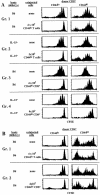
Homeostatic proliferation of naive and memory T cells in T cell–depleted syngenic hosts. (A) LN and spleen cells from B6.PL mice were sorted for naive (CD44lo) or memory phenotype (CD44hi) CD4+ and CD8+ T cells. Small numbers (106 cells per mouse) of CFSE-labeled CD44lo or CD44hi cells were intravenously injected into irradiated (600 cGy) B6, B6. H2-Aβ, and β2mKD mice; proliferation of donor cells was analyzed 7 d later by flow cytometry after staining host LN and spleen cells for Thy-1.1 and CD4. Shown are CD44 profiles on the sorted donor cells before injection (left column), and the CFSE profiles on gated donor CD4+ (Thy-1.1+ CD4+) and CD8+ (Thy-1.1+ CD4) cells in the LN in the indicated hosts (right columns). Similar results were found in the spleen. (B) Small numbers (106 cells per mouse) of sorted CFSE-labeled CD44lo or CD44hi T cells were injected into two groups of irradiated (600 cGy) B6, B6.β2m, B6.Aβ, and B6.β2m mice; proliferation of donor cells was analyzed 7 d later. The injected donor cells were deficient in MHC class I molecules to prevent rejection by B6.β2m mice. MHC class I–deficient donor cells were obtained from mixed BM chimeras generated by reconstituting lethally irradiated B6 mice with a mixture of β2m-deficient B6.Ly 5.1+ and normal B6 BM cells as described in Materials and Methods. Shown are representative CFSE profiles on gated donor CD44lo or CD44hi CD4+ (Ly5.1+ CD4+) cells in the host LN. All host mice were pretreated with anti-NK1.1 (clone PK136) mAb to prevent rejection of β2m donor cells. Similar results were obtained from two other experiments.
Figure 1.
Figure 1.
Homeostatic proliferation of naive and memory T cells in T cell–depleted syngenic hosts. (A) LN and spleen cells from B6.PL mice were sorted for naive (CD44lo) or memory phenotype (CD44hi) CD4+ and CD8+ T cells. Small numbers (106 cells per mouse) of CFSE-labeled CD44lo or CD44hi cells were intravenously injected into irradiated (600 cGy) B6, B6. H2-Aβ, and β2mKD mice; proliferation of donor cells was analyzed 7 d later by flow cytometry after staining host LN and spleen cells for Thy-1.1 and CD4. Shown are CD44 profiles on the sorted donor cells before injection (left column), and the CFSE profiles on gated donor CD4+ (Thy-1.1+ CD4+) and CD8+ (Thy-1.1+ CD4) cells in the LN in the indicated hosts (right columns). Similar results were found in the spleen. (B) Small numbers (106 cells per mouse) of sorted CFSE-labeled CD44lo or CD44hi T cells were injected into two groups of irradiated (600 cGy) B6, B6.β2m, B6.Aβ, and B6.β2m mice; proliferation of donor cells was analyzed 7 d later. The injected donor cells were deficient in MHC class I molecules to prevent rejection by B6.β2m mice. MHC class I–deficient donor cells were obtained from mixed BM chimeras generated by reconstituting lethally irradiated B6 mice with a mixture of β2m-deficient B6.Ly 5.1+ and normal B6 BM cells as described in Materials and Methods. Shown are representative CFSE profiles on gated donor CD44lo or CD44hi CD4+ (Ly5.1+ CD4+) cells in the host LN. All host mice were pretreated with anti-NK1.1 (clone PK136) mAb to prevent rejection of β2m donor cells. Similar results were obtained from two other experiments.
Figure 2.
Figure 2.
Homeostatic proliferation of naive and memory phenotype T cells in cytokine-deficient mice. Small numbers (106 cells per mouse) of CFSE-labeled sorted B6.Ly 5.1 CD44lo, and CD44hi T cells were injected into groups of irradiated (600 cGy) B6, B6.IL-4, IL-7, and B6.IL-15 mice and analyzed 7 d later for proliferation of donor cells in host lymphoid tissues. Shown are CFSE profiles of gated donor CD44lo or CD44hi CD4+ (Ly5.1+ CD4+) and CD8+ (Ly5.1+ CD4) cells in host LN 7 d after transfer. Data are representative of two separate experiments using a total of four mice for each host type.
Figure 3.
Figure 3.
Role of IL-7 and IL-15 for homeostatic proliferation of naive and memory phenotype T cells. (A) Expression of IL-2Rβ (CD122), IL-7Rα (CD127), and γc (CD132) on CD44lo or CD44hi T cells. LN cells from a young B6.PL mouse were stained for the indicated cytokine receptors and also for CD44, CD4, and CD8. Shown are expression levels of the indicated cytokine receptors (black) and background control (white) on gated CD44lo, CD44hi CD4+, and CD8+ cells. (B) Blocking IL-7 in IL-15 mice suppresses homeostatic proliferation of CD44hi CD8+ cells. A mixture of CFSE-labeled sorted B6.Ly 5.1+ CD44lo T cells (106 cells per mouse) and B6.PL CD44hi T cells (106 cells per mouse) were injected into irradiated (600 cGy) B6, IL-7, and IL-15 mice. Host mice were either treated with a mixture of control mouse and rat IgG or with a cocktail of anti–IL-7Rα (clone A7R34) and anti–IL-7 (clone M25) mAbs. Cocktails containing 500 ug of each antibody were intraperitoneally injected every other day for a total of four times starting 1 d before cell transfer. Shown are CFSE profiles of donor CD44loCD4+ (Ly 5.1+ CD4+), CD44lo CD8+ (Ly5.1+ CD4), CD44hi CD4+ (Thy1.1+ CD4+), or CD44hi CD8+ (Thy1.1+ CD4) cells in the host LN 7 d after transfer. One other experiment showed similar results.
Figure 4.
Figure 4.
Mice deficient in both IL-7 and IL-15 fail to support homeostatic proliferation of memory phenotype CD8+ cells but support efficient homeostatic proliferation of memory phenotype CD4+ cells. Small numbers (106) of sorted and CFSE-labeled B6.PL CD44hi T cells were transferred into irradiated B6, IL-7, IL-15, and combined IL-7 IL-15 mice; proliferation of donor cells was examined 7 d later. Shown are CFSE profiles of gated donor CD44hi CD4+ and CD44hi CD8+ cells in the LN. Similar results were obtained from two other experiments.
Figure 5.
Figure 5.
Competition between naive and memory phenotype T cells for factors driving homeostatic proliferation. (A) Coinjection of large numbers of CD44lo T cells fails to inhibit homeostatic proliferation of CD44hi CD8+ cells, but coinjection of large numbers of CD44hi CD8+ cells blocks homeostatic proliferation of CD44lo T cells. A mixture of CFSE-labeled sorted B6.Ly 5.1+ CD44lo T cells (106 cells per mouse) and B6.PL CD44hi T cells (106 cells per mouse) was injected into a group of irradiated (600 cGy) B6 or B6.IL-15 mice. Half of the hosts were then injected with a large number (3–4 × 10⁷) of magnetic bead-purified B6 CD44lo T cells or CD44hi CD8+ cells obtained as described in Materials and Methods. CD44hi CD8+ cells were obtained from B6.IL-7 transgenic mice. Donor cells in the host LN and spleen were examined 7 d later. Shown are CFSE profiles on gated donor CD44lo and CD44hi CD8+ cells. Note that each group represents separate experiments; hence, representative control proliferations of CFSE-labeled donor cells in the absence of coinjected bystander cells is shown for each group. Results are representative of two to three independent experiments. (B) Failure of bystander naive T cells or CD44hi CD8+ cells to diminish proliferation of CD44hi CD4+ cells. Representative data on gated donor CD44lo and CD44hi CD4+ cells from some of the experiments described in A are shown.

"VSports手机版" Comment in

References

    1. Freitas, A.A., and B.B. Rocha. 1993. Lymphocyte lifespan: homeostasis, selection and competition. Immunol. Today. 14:25–29. - PubMed
    1. Bell, E.B., and S.M. Sparshott. 1997. The peripheral T-cell pool: regulation by non-antigen induced proliferation? Sem. Immunol. 9:347–353. - PubMed
    1. Goldrath, A.W., and M.J. Bevan. 1999. Selecting and maintaining a diverse T-cell repertoire. Nature. 402:255–262. - PubMed
    1. Surh, C.D., and J. Sprent. 2000. Homeostatic T cell proliferation. How far can T cells be activated to self-ligands? J. Exp. Med. 192:F9–F14. - PMC - PubMed
    1. Viret, C., F.S. Wong, and C.A. Janeway, Jr. 1999. Designing and maintaining the mature TCR repertoire: the continuum of self-peptide:self-MHC complex recognition. Immunity. 10:559–568. - PubMed

"V体育官网" Publication types

"V体育2025版" MeSH terms