"V体育官网" Skip to main page content
U.S. flag

An official website of the United States government

Dot gov

The . gov means it’s official. Federal government websites often end in . gov or . mil VSports app下载. Before sharing sensitive information, make sure you’re on a federal government site. .

Https

The site is secure V体育官网. The https:// ensures that you are connecting to the official website and that any information you provide is encrypted and transmitted securely. .

Comparative Study
. 2002 Jun;13(6):1929-39.
doi: 10.1091/mbc.02-02-0023.

"VSports注册入口" Gene expression patterns in human liver cancers

Affiliations
Comparative Study

Gene expression patterns in human liver cancers

VSports手机版 - Xin Chen et al. Mol Biol Cell. 2002 Jun.

Abstract

Hepatocellular carcinoma (HCC) is a leading cause of death worldwide. Using cDNA microarrays to characterize patterns of gene expression in HCC, we found consistent differences between the expression patterns in HCC compared with those seen in nontumor liver tissues. The expression patterns in HCC were also readily distinguished from those associated with tumors metastatic to liver. The global gene expression patterns intrinsic to each tumor were sufficiently distinctive that multiple tumor nodules from the same patient could usually be recognized and distinguished from all the others in the large sample set on the basis of their gene expression patterns alone VSports手机版. The distinctive gene expression patterns are characteristic of the tumors and not the patient; the expression programs seen in clonally independent tumor nodules in the same patient were no more similar than those in tumors from different patients. Moreover, clonally related tumor masses that showed distinct expression profiles were also distinguished by genotypic differences. Some features of the gene expression patterns were associated with specific phenotypic and genotypic characteristics of the tumors, including growth rate, vascular invasion, and p53 overexpression. .


Figures
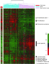
Figure 1
Figure 1
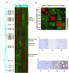
Hierarchical clustering of the patterns of variation in expression of 3180 genes (represented by 3964 cDNA), in 156 liver tissues (74 nontumor liver and 82 HCC). The data are shown in a table format, in which rows represent individual genes and columns represent individual tissue or cell sample. The color in each cell reflects the expression level of the corresponding gene in the corresponding tissue, relative to its mean expression level across the entire set of tissue samples. The scale extends from fluorescence ratios of 0.25–4 relative to the mean level for all samples. Gray indicates missing or excluded data. Expression of the same genes in 10 HCC cell lines is similarly represented in the panel to the left of the main panel. See supplementary information for the full data, including sample names.
Figure 2
Figure 2
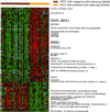
Features of the variation in gene expression patterns can be related to specific physiological or histological features of the samples. Data are the same as in Figure 1. Proliferation cluster (A), liver-specific cluster (B), T-lymphocytes cluster (C), B-lymphocytes cluster (D), stromal cell cluster 1 (E), stromal cell cluster 2 (F), endothelial cell cluster 1 (G); endothelial cell cluster 2 (H), immunohistochemistry staining of CD34 on nontumor liver (upper) and HCC (lower) tissues (I). Due to limited space, only a few selected gene names are shown. Only a portion of the proliferation cluster and the liver-specific cluster are shown. See supplementary information for the full data.
Figure 3
Figure 3
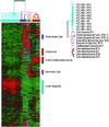
Gene expression profiles in HCC samples. (A) Hierarchical clustering of 3271 clones representing ∼2640 different genes and 102 HCC samples (from 82 patients) based on similarity in gene expression patterns. The scale is the same as Figure 1. The dendrogram on the left shows the samples analyzed. Branches are colored to highlight groups of samples from the same patient. Each blue line represents a patient from whom only a single tumor sample was analyzed. Each of the other colors represents multiple tumor samples from a single patient. (B) r values between multiple samples. Using all the genes for which technically adequate measurements could be obtained, we calculated the r values for the expression patterns of each pair of samples. In this image, each cell represents the r for one pair of samples, using the color key indicated to the right of the panel. (C) Immunohistochemical staining of tumors staining HK65 T1, T2, and T4 for p53. (D) Immunohistochemical staining of tumors HK67 T1 (left two) and T2 and T3 (right two) for p53.
Figure 4
Figure 4
Hierarchical clustering of 121 genes whose expression level was significantly correlated with the presence or absence of nuclear p53 immunostaining in HCC (permutation p value < 0.001; corresponding to a PFER of 4.043 and a nominal FDR of ∼0.035). The scale is the same as Figure 1. The colored bars above the panel indicate p53 positive (yellow) or negative tumor samples (brown). Due to space limitation, only some of the genes are labeled. The complete data with clone ID and gene names are available at the supplementary information.
Figure 5
Figure 5
Hierarchical clustering of 91 genes whose expression was significantly correlated with vascular invasion in HCC (permutation p value < 0.001; corresponding to a PFER of 4.043 and a nominal FDR of ∼0.042). The scale is the same as Figure 1. The colored bars above the panel indicate tumors with vascular invasion (purple) or without vascular invasion (orange). See supplementary information for full data.
Figure 6
Figure 6
Comparison of gene expression patterns in HCC and tumors metastatic to liver. Expression data for 3474 cDNA clones representing ∼2780 different genes, in 10 HCC and 10 metastatic tumor samples, were analyzed by hierarchical clustering. The scale is the same as Figure 1. Differently colored branches in the dendrogram represent different histologically defined tumor types. See supplementary information for full data.

References

    1. Alizadeh AA, et al. Distinct types of diffuse large B-cell lymphoma identified by gene expression profiling. Nature. 2000;403:503–511. - PubMed
    1. Arii S, Mise M, Harada T, Furutani M, Ishigami S, Niwano M, Mizumoto M, Fukumoto M, Imamura M. Overexpression of matrix metalloproteinase 9 gene in hepatocellular carcinoma with invasive potential. Hepatology. 1996;24:316–322. - PubMed
    1. Beasley RP. Hepatitis B virus. The major etiology of hepatocellular carcinoma. Cancer. 1988;61:1942–1956. - PubMed
    1. Caruso ML, Valentini AM. Overexpression of p53 in a large series of patients with hepatocellular carcinoma: a clinicopathological correlation. Anticancer Res. 1999;19:3853–3856. - PubMed (VSports在线直播)
    1. Cho RJ, Huang M, Dong H, Steinmetz L, Sapinoso L, Hampton G, Elledge SJ, Davis RW, Lockhart DJ, Campbell MJ. Transcriptional regulation and function during the human cell cycle. Nat Genet. 2001;27:48–54. - PubMed (V体育官网)

Publication types

VSports手机版 - MeSH terms

Substances (V体育ios版)