Skip to main page content
U.S. flag

An official website of the United States government

Dot gov

The . gov means it’s official VSports app下载. Federal government websites often end in . gov or . mil. Before sharing sensitive information, make sure you’re on a federal government site. .

Https

The site is secure. The https:// ensures that you are connecting to the official website and that any information you provide is encrypted and transmitted securely V体育官网. .

Case Reports
. 2002 Jun;109(11):1501-9.
doi: 10.1172/JCI14858.

Deficient natural killer cell cytotoxicity in patients with IKK-gamma/NEMO mutations

Affiliations
Case Reports

Deficient natural killer cell cytotoxicity in patients with IKK-gamma/NEMO mutations

V体育官网入口 - Jordan S Orange et al. J Clin Invest. 2002 Jun.

Abstract

NF-kappaB essential modifier (NEMO), also known as IKK-gamma, is a member of the I-kappaB kinase complex responsible for phosphorylating I-kappaB, allowing the release and activation of NF-kappaB. Boys with an expressed NEMO mutation have an X-linked syndrome characterized by hypohidrotic ectodermal dysplasia with immune deficiency (HED-ID). The immunophenotype resulting from NEMO mutation is highly variable, with deficits in both T and B cell responses. We evaluated three patients with NEMO mutations (L153R, Q403X, and C417R) and HED-ID who had evidence of defective CD40 signaling. All three patients had normal percentages of peripheral blood NK cells, but impaired NK cell cytotoxic activity. This was not due to a generalized defect in cytotoxicity because antibody-dependent cellular cytotoxicity was intact. This abnormality was partially reversed by in vitro addition of IL-2, which was also able to induce NF-kappaB activation VSports手机版. In one patient with recurrent cytomegalovirus infections, administration of IL-2 partially corrected the NK cell killing deficit. These data suggest that NEMO participates in signaling pathways leading to NK cell cytotoxicity and that IL-2 can activate NF-kappaB and partially overcome the NK cell defect in patients with NEMO mutations. .


Figures (VSports)

Figure 1
Figure 1
NEMO mutations in patients with HED-ID. A schematic diagram of the human NEMO protein is shown with the individual domains labeled in boxes (αH, α-helix; C-C, coiled-coil; LZ, leucine zipper; ZF, zinc finger). Sequences containing amino acid substitutions in the three patients studied are displayed in brackets below the gene map. Arrows indicate positions of amino acid substitutions. The particular amino acid altered (underlined) and the substituted residues (circled) are shown. The specific nucleotide point mutation resulting in the missense amino acid is listed above the gene mutation.
Figure 2
Figure 2
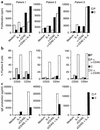
Functional consequences of CD40 ligation in PBMC cultures from patients with NEMO mutation. (a) Proliferation of PBMCs incubated with control medium, IL-4, anti-CD40, or IL-4 + anti-CD40 was determined by 3H incorporation. Black bars show proliferation in a control donor and white bars show proliferation in patients. Data are representative of two experiments each from each patient. (b) Surface expression of CD23 and CD54 was evaluated on CD20+ lymphocytes in representative controls (black bars) or patients (dark gray bars) and on anti-CD40–stimulated cells of controls (white bars) or patients (light gray bars). (c) IgE production was measured in supernatants of PBMC cultures from control donors (black bars) or patients (white bars) after incubation with medium, sCD40L, IL-4, or sCD40L + IL-4.
Figure 3
Figure 3
Evaluation of nuclear NF-κB level in cells from a patient with NEMO exon 4 mutation (patient 1). Nuclear extracts of enriched B cells from patient 1 or a control donor were evaluated by EMSA using a 32P-labeled NF-κB probe. (a) The effect of CD40 ligation on the level of nuclear NF-κB was determined by incubation of enriched B cells with control medium or sCD40L for 30 minutes prior to cell lysis. (b) The ability of IL-2 to induce nuclear levels of NF-κB was assessed in PBMCs by stimulation with PHA for 18 hours, followed by resting for 5 hours and subsequent incubation with IL-2 or media for 5 hours prior to lysis. Nuclear extracts were prepared and evaluated for NF-κB binding. The arrow points to the retained NF-κB probe; the large band at the bottom of the gel corresponds to free probe.
Figure 4
Figure 4
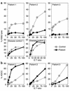
Deficient NK cell cytotoxicity in patients with HED-ID and NEMO mutation. (a) NK cell cytotoxicity was assessed in PBMCs from patients (circles) and controls (squares) by 51Cr-release assay with K562 target cells (top panels). Results are representative of a total of five experiments from patient 1 and two experiments each from patients 2 and 3. Patient and control samples were evaluated in parallel. Disease controls (bottom panels) are as described in Methods. (b) ADCC was measured in patients (circles) and controls (squares) using CL27A cells and antiserum (patients 1 and 3), or ZKBB cells and anti-CD20 monoclonal antibody (patient 2) as target cells. Lysis of 51Cr-labeled target cells was determined after 4 hours. Results are representative of three individual experiments for patient 1 and one experiment for patients 2 and 3. Lysis of target cells without added antibody was not detected. ADCC was always evaluated in parallel with K562 lysis.
Figure 5
Figure 5
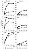
In vitro IL-2 induction of NK cell cytotoxicity in PBMCs of patients with HED-ID and NEMO mutation. The effect of IL-2 on NK cell cytotoxic activity by PBMCs from control donors and patients was evaluated in parallel. Effector cells were combined with 51Cr-labeled target cells without (circles) or with 1,000 U/ml human recombinant IL-2 added for the 4-hour duration of the assay (diamonds). Results are representative of three individual experiments for patient 1 and two each for patients 2 and 3.
Figure 6
Figure 6
Induction of ex vivo NK cell cytotoxic activity after in vivo administration of IL-2 to a patient with NEMO exon 4 mutation. Patient 1 was treated with 1 × 10⁶ U/m2 of human recombinant IL-2 by continuous intravenous infusion for 5 days. NK cytotoxicity was determined ex vivo by lysis of 51Cr-labeled target cells before treatment, immediately after (0 wk), 1 week after, 2 weeks after, 3 weeks after, or 4 weeks after treatment (circles). K562 lysis mediated by PBMCs from untreated control donors (squares) was performed in parallel. Different control donors were used in each assay.

References

    1. Clarke A, Phillips DI, Brown R, Harper PS. Clinical aspects of X-linked hypohidrotic ectodermal dysplasia. Arch Dis Child. 1987;62:989–996. - PMC - PubMed
    1. Kere J, et al. X-linked anhidrotic (hypohidrotic) ectodermal dysplasia is caused by mutation in a novel transmembrane protein. Nat Genet. 1996;13:409–416. - PubMed
    1. Monreal AW, et al. Mutations in the human homologue of mouse dl cause autosomal recessive and dominant hypohidrotic ectodermal dysplasia. Nat Genet. 1999;22:366–369. - PubMed
    1. Headon DJ, et al. Gene defect in ectodermal dysplasia implicates a death domain adapter in development. Nature. 2001;414:913–916. - PubMed (VSports手机版)
    1. Abinun M, Spickett G, Appleton AL, Flood T, Cant AJ. Anhidrotic ectodermal dysplasia associated with specific antibody deficiency. Eur J Pediatr. 1996;155:146–147. - PubMed

Publication types

"VSports" MeSH terms