Skip to main page content
U.S. flag

An official website of the United States government

Dot gov

The VSports app下载. gov means it’s official. Federal government websites often end in . gov or . mil. Before sharing sensitive information, make sure you’re on a federal government site. .

Https

The site is secure V体育官网. The https:// ensures that you are connecting to the official website and that any information you provide is encrypted and transmitted securely. .

. 2002 May 1;16(9):1089-101.
doi: 10.1101/gad.988402.

The Dlx5 and Dlx6 homeobox genes are essential for craniofacial, axial, and appendicular skeletal development

Affiliations

The Dlx5 and Dlx6 homeobox genes are essential for craniofacial, axial, and appendicular skeletal development

Raymond F Robledo et al. Genes Dev. .

Abstract

Dlx homeobox genes are mammalian homologs of the Drosophila Distal-less (Dll) gene. The Dlx/Dll gene family is of ancient origin and appears to play a role in appendage development in essentially all species in which it has been identified. In Drosophila, Dll is expressed in the distal portion of the developing appendages and is critical for the development of distal structures. In addition, human Dlx5 and Dlx6 homeobox genes have been identified as possible candidate genes for the autosomal dominant form of the split-hand/split-foot malformation (SHFM), a heterogeneous limb disorder characterized by missing central digits and claw-like distal extremities. Targeted inactivation of Dlx5 and Dlx6 genes in mice results in severe craniofacial, axial, and appendicular skeletal abnormalities, leading to perinatal lethality. For the first time, Dlx/Dll gene products are shown to be critical regulators of mammalian limb development, as combined loss-of-function mutations phenocopy SHFM. Furthermore, spatiotemporal-specific transgenic overexpression of Dlx5, in the apical ectodermal ridge of Dlx5/6 null mice can fully rescue Dlx/Dll function in limb outgrowth. VSports手机版.


V体育官网 - Figures
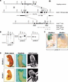
Figure 1
Figure 1
Simultaneous disruption of Dlx5 and Dlx6 by homologous recombination, genotyping, mRNA, and morphological analyses of embryos carrying a Dlx5/6 null allele. (A) Structure of the genomic Dlx5 and Dlx6 loci, ires.lacZ.neo targeting vector, and mutant allele following homologous recombination. Exons are depicted as black boxes. The transcriptional orientation of each gene is indicated. The probes employed for Southern and RNA in situ analyses are indicated with black rectangles. (B) Southern blot analysis of embryonic genomic DNA isolated from the yolk-sac was digested with BamHI and then hybridized with a 5′ (probe 1) in the undeleted region of Dlx5, 5′ (probe 2) in the undeleted region of Dlx6, or 3′ (probe 3) in the deleted region of Dlx5. The wild-type and mutant alleles were detected as 6.6 kb and 2.2 kb fragments, respectively, with probe 1 and 4.3 kb and 4.6 kb, respectively, with probe 2. The wild-type allele was detected as a 6.6 kb fragment and the mutant allele was undetected with probe 3. (C) Whole-mount in situ hybridization of E10.5 wild-type and Dlx5/6−/− embryos using probe 4. Dlx5/6−/− embryos lack probe 4 expression as probe 4 falls within the region deleted in the Dlx5/6−/− embryos. (D) Gross appearance of wild-type and Dlx5/6−/− embryos and hindlimbs at E18.5. Dlx5/6−/− embryos show an overall reduced size, exencephaly, kinked tail vertebrae, and split hindlimbs. Scanning electron microscopy (SEM) of whole-mount wild-type and Dlx5/6−/− embryos at E12.5, highlighting the craniofacial, tail, and limb malformations already clearly visible at this stage. Abbreviations: aer, apical ectodermal ridge; ba, branchial arches; ov, otic vesicle.
Figure 2
Figure 2
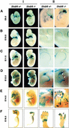
Embryonic Dlx5 and Dlx6 expression revealed by whole-mount β-galactosidase staining of heterozygous and homozygous Dlx5/6 mutant embryos (Column I), with higher magnifications of limbs from the same embryos (Column II). (A) Dlx5/6 is strongly expressed in the first and second branchial arches and the otic pit, with less prominent expression in the tail and forelimb buds at E9.5. (B) Expression becomes apparent in the AER of both fore- and hindlimb buds by E10.5. (C) At E11.5, reduction in expression in the frontonasal prominence and medial AER (arrow) initiates. (D) Expression in the medial AER deteriorates by E12.5 in Dlx5/6−/− embryos. Arrows in the right-hand column indicate the loss of medial tissue and digits in the Dlx5/6−/− embryos. (E) Expression within all developing bones becomes apparent by E14.5, at which time the SHFM phenotype becomes recognizable in null mutants. (F) At E18.5, craniofacial and skeletal expression remains strong. Calvarial and mandibular expression is noticeably lost in Dlx5/6−/− embryos owing to the absence of these tissues.
Figure 3
Figure 3
Craniofacial, axial, and appendicular skeletal defects of Dlx5/6−/− embryos. (A) Whole-mount Alcian blue (AB) staining reveals the absence of Meckel's, nasal prominence and central digit cartilages of E14.5 mutant embryos. (B) Alcian blue and alizarin red (AB/AR) staining shows the absence of calvaria, maxillary, and mandibular bones of E16.5 mutant embryos. In addition, the axial skeleton, including vertebral bodies (inset), has little or no ossification and the ribs are malformed. The forelimbs (left columns) also have a delay in ossification (bracketed region), or a complete absence of ossification (star) and hindlimb digits (right columns) are absent and/or fused. (C) The delay in ossification becomes less severe in the axial and appendicular skeleton by E18.5. However, the ribs remain disfigured and the ratio of forelimb ossification to cartilage remains retarded. Hindlimbs and forelimbs (inset) have clefting due to missing and fused digits. Abbreviations: C, calvaria; FN, frontonasal cartilage; MC, Meckel's cartilage; Md; Mandible.
Figure 4
Figure 4
Histological and molecular examination of the ossification of the scapula of heterozygous and Dlx5/6 null embryos at E16.5. (A) AB/AR staining of whole-mount scapulas shows the absence of ossification of the spine of scapula (Ss) and a minimal ossification within the center of the scapula, compared to heterozygous embryos. Vertical lines indicate the approximate area from which comparable serial sections were taken for each column of results. (B) In more proximal sections, AB-stained Dlx5/6−/− scapulas appear to lack both hypertrophic chondrocytes and a von Kossa (VK)-positive calcium matrix. (C) In contrast to control embryos, Col2a1 expression remains strong and Col10a1 appears to be increasing in the Dlx5/6 null sections. In addition, Ihh downregulation appears to be delayed in Dlx5/6 null sections, whereas Pthrp-r expression is unaffected. Runx2 expression appears normal in the perichondrium, but reduced in the chondrium of the Dlx5/6 null sections. Osteocalcin expression is completely absent in the Dlx5/6 null sections. In more distal sections, calcified hypertrophic chondrocytes are now apparent. Col2a1 expression is now almost absent in the Dlx5/6 null sections, and Col10a1 expression is strong. At this level, Runx2 appears normal in the Dlx5/6 null sections and Osteocalcin expression remains absent.
Figure 5
Figure 5
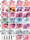
Molecular analysis of altered limb development of Dlx5/6−/− embryos. Whole-mount in situ hybridization shows that Shh and Lmx1b expression is normal at E10.5 and E11.5 in the Dlx5/6 null embryos. Fgf8 and Msx2 expression in the hindlimb AER is normal at E10.5, but is absent in the medial AER by E11.5. Arrowheads in the right-hand column indicate the loss of expression and missing tissue in the medial portion of the distal limb in the Dlx5/6−/− embryos. Analysis with a Dlx5 5′ riboprobe for the Dlx5 exon still present in the mutant allele reveals diminished Dlx5 expression in the medial AER by E11.5. Immunohistochemical staining and quantitation of BrdU incorporation reveals decreased cellular proliferation in the AER at E10.5 and E11.5, while proliferation in the underlying mesenchyme is unaffected. A compensatory increase of Dlx2 expression in the AER was not observed.
Figure 6
Figure 6
SHFM phenotypic rescue of Dlx5/6−/− embryos by Dlx5 transgene expression in the developing AER. (A) Structure of the Dlx5 and alkaline phosphatase (AP) transgenes that employ the Msx2 AER-specific enhancer. (B) Whole-mount in situ hybridization of AER-Dlx5/AP embryos showing AER expression of the tagged (TAG) Dlx5 transgene and transgene-specific alkaline phosphatase staining of the AER at E10.5. (C) AB staining at E14.5 shows restored hindlimb cartilage outgrowth and patterning of Dlx5/6−/− embryos expressing the AER-Dlx5/AP transgene. AB/AR cartilage and bone staining of the hindlimbs remains rescued at E18.5. Whole-mount in situ hybridization at E12.5 shows that AER expression of both Fgf8 and Msx2 can be restored by AER-Dlx5/AP expression in mutant mice. Arrowheads at E12.5 indicate the loss of marker expression and missing tissue in the medial portion of the distal limb in the Dlx5/6−/− embryos lacking the AER-Dlx5/AP transgene.
Figure 6
Figure 6
SHFM phenotypic rescue of Dlx5/6−/− embryos by Dlx5 transgene expression in the developing AER. (A) Structure of the Dlx5 and alkaline phosphatase (AP) transgenes that employ the Msx2 AER-specific enhancer. (B) Whole-mount in situ hybridization of AER-Dlx5/AP embryos showing AER expression of the tagged (TAG) Dlx5 transgene and transgene-specific alkaline phosphatase staining of the AER at E10.5. (C) AB staining at E14.5 shows restored hindlimb cartilage outgrowth and patterning of Dlx5/6−/− embryos expressing the AER-Dlx5/AP transgene. AB/AR cartilage and bone staining of the hindlimbs remains rescued at E18.5. Whole-mount in situ hybridization at E12.5 shows that AER expression of both Fgf8 and Msx2 can be restored by AER-Dlx5/AP expression in mutant mice. Arrowheads at E12.5 indicate the loss of marker expression and missing tissue in the medial portion of the distal limb in the Dlx5/6−/− embryos lacking the AER-Dlx5/AP transgene.

References

    1. Acampora D, Merlo GR, Paleari L, Zerega B, Postiglione MP, Mantero S, Bober E, Barbieri O, Simeone A, Levi G. Craniofacial, vestibular and bone defects in mice lacking the Distal-less-related gene Dlx5. Development. 1999;126:3795–3809. - PubMed
    1. Ahuja HS, Zhu Y, Zakeri Z. Association of cyclin-dependent kinase 5 and its activator p35 with apoptotic cell death. Dev Genet. 1997;21:258–267. - "VSports app下载" PubMed
    1. Capdevila J, Izpisua Belmonte JC. Patterning mechanisms controlling vertebrate limb development. Annu Rev Cell Dev Biol. 2001;17:87–132. - PubMed
    1. Chai CK. Dactylaplasia in mice a two-locus model for development anomalies. J Hered. 1981;72:234–237. - PubMed (V体育平台登录)
    1. Charite J, McFadden DG, Merlo G, Levi G, Clouthier DE, Yanagisawa M, Richardson JA, Olson EN. Role of Dlx6 in regulation of an endothelin-1-dependent, dHAND branchial arch enhancer. Genes Dev. 2001;15:3039–3049. - "VSports注册入口" PMC - PubMed

Publication types

MeSH terms