"V体育官网" Skip to main page content
U.S. flag

An official website of the United States government

Dot gov

The . gov means it’s official. Federal government websites often end in . gov or . mil. Before sharing sensitive information, make sure you’re on a federal government site. VSports app下载.

Https

The site is secure. The https:// ensures that you are connecting to the official website and that any information you provide is encrypted and transmitted securely V体育官网. .

. 2002 Apr 1;195(7):801-10.
doi: 10.1084/jem.20011481.

"V体育平台登录" c-Jun NH(2)-terminal kinase (JNK)1 and JNK2 signaling pathways have divergent roles in CD8(+) T cell-mediated antiviral immunity

Affiliations

c-Jun NH(2)-terminal kinase (JNK)1 and JNK2 signaling pathways have divergent roles in CD8(+) T cell-mediated antiviral immunity

Nathalie Arbour et al. J Exp Med. .

Abstract

c-Jun NH(2)-terminal kinases (JNK) play important roles in T helper cell (Th) proliferation, differentiation, and maintenance of Th1/Th2 polarization. To determine whether JNKs are involved in antiviral T cell immunity, and whether JNK1 and JNK2 bear biological differences, we investigated the immune responses of JNK1-deficient and JNK2-deficient mice to lymphocytic choriomeningitis virus (LCMV). After LCMV infection, wild-type (JNK(+/+)) mice had a 5- to 10-fold increase in splenic CD8(+) T cells. In contrast, infected JNK1(-/-) mice showed a significantly lower virus-specific CD8(+) T cell expansion. However, JNK1(-/-) mice cleared LCMV infection with similar kinetics as JNK(+/+) mice VSports手机版. Splenic T cells from LCMV-infected JNK1(-/-) animals produced interferon gamma after stimulation with viral peptides. However, fewer JNK1(-/-) T cells acquired an activated phenotype (CD44(hi)) and more JNK1(-/-)CD8(+)CD44(hi) cells underwent apoptosis than JNK(+/+) cells at the peak of the primary response. In contrast, LCMV-infected JNK2(-/-) mice generated more virus-specific CD8(+) T cells than JNK(+/+) mice. These results indicate that JNK1 and JNK2 signal pathways have distinct roles in T cell responses during a viral infection. JNK1 is involved in survival of activated T cells during immune responses, and JNK2 plays a role in control of CD8(+) T cell expansion in vivo. .


Figures

Figure 1.
Figure 1.
After LCMV infection, the percentage of virus specific splenic T cells is higher in JNK2−/−, but similar in JNK1−/− and JNK+/+ mice. JNK1−/−, JNK2−/−, and JNK+/+ animals were injected intraperitoneally with LCMV ARM 2 × 10⁵ PFU. 7 (D7) or 8 (D8) d after infection, spleens were processed and stimulated for 5 h in vitro with virus specific peptide GP aa 33–41. Cells were then stained for CD8 and intra-cellular IFN-γ. Values written in the dot plots indicate the percentage of CD8+ T cells positive for IFN-γ.
Figure 2.
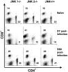
Figure 2.
The CD8+ T cell compartment does not expand in JNK1−/− mice during the primary LCMV infection in comparison to JNK2−/− and JNK+/+ mice but equivalent number of CD8+ T cell are noted 60 d later. JNK+/+, JNK1−/−, and JNK2−/− mice were injected intraperitoneally with LCMV ARM 2 × 10⁵ PFU. Uninfected spleen cells and spleen cells 7 (D7) or 60 d (D60) postinfection were stained for CD4 and CD8. Data represent average values obtained from three animals per group and are representative of two independent experiments. Values written in the dot plots indicate the percentage of CD8+ T cells and the percentage of CD4+ T cells
Figure 3.
Figure 3.
The number of virus-specific CD8+ T cells and CD4+ T cells is lower in JNK1−/− mice than in either JNK2−/− or JNK+/+ mice after LCMV infection. JNK1−/−, JNK2−/−, and JNK+/+ mice were injected intraperitoneally with LCMV ARM, 2 × 10⁵ PFU. 8 d after infection, spleen were processed and stimulated for 5 h in vitro with virus-specific peptide GP aa 33–41, NP aa 396–404, GP aa 61–80, or NP aa 309–328. Cells were then stained for surface CD4 or CD8 and intracellular IFN-γ molecules. The number of virus-specific T cells was calculated from the number of total spleen cells obtained per animal. Data represent average values obtained from three animals per group and are representative of two independent experiments. (A) CD8+ T cells; (B) CD4+ T cells. *P < 0.05 #P = 0.052 when compared with JNK+/+.
Figure 3.
Figure 3.
The number of virus-specific CD8+ T cells and CD4+ T cells is lower in JNK1−/− mice than in either JNK2−/− or JNK+/+ mice after LCMV infection. JNK1−/−, JNK2−/−, and JNK+/+ mice were injected intraperitoneally with LCMV ARM, 2 × 10⁵ PFU. 8 d after infection, spleen were processed and stimulated for 5 h in vitro with virus-specific peptide GP aa 33–41, NP aa 396–404, GP aa 61–80, or NP aa 309–328. Cells were then stained for surface CD4 or CD8 and intracellular IFN-γ molecules. The number of virus-specific T cells was calculated from the number of total spleen cells obtained per animal. Data represent average values obtained from three animals per group and are representative of two independent experiments. (A) CD8+ T cells; (B) CD4+ T cells. *P < 0.05 #P = 0.052 when compared with JNK+/+.
Figure 4.
Figure 4.
During the acute infection, the number of splenic CD8+ T cells CD44hi is lower in JNK1−/− mice than in JNK2−/− and JNK+/+ mice, and a greater proportion of these cells from JNK1−/− undergo apoptosis. JNK1−/−, JNK2−/−, and JNK+/+ animals were injected intraperitoneally with LCMV ARM 2 × 10⁵ PFU. 8 d after infection, spleen were processed to make single cell suspension devoted of red blood cells and stained directly for surface markers: CD4, CD8, and CD44 then annexin-V and 7AA-D were added. Data represent average values obtained from four animals per group and are representative of two independent experiments. The percentage of CD8+ or CD4+ that were CD44hi are shown in the left, that were annexin V+ cells among the CD8CD44hi or CD4CD44hi on the right. (A) CD8+ T cells. (B) CD4+ T cells. *P < 0.05 compared with JNK+/+.
Figure 4.
Figure 4.
During the acute infection, the number of splenic CD8+ T cells CD44hi is lower in JNK1−/− mice than in JNK2−/− and JNK+/+ mice, and a greater proportion of these cells from JNK1−/− undergo apoptosis. JNK1−/−, JNK2−/−, and JNK+/+ animals were injected intraperitoneally with LCMV ARM 2 × 10⁵ PFU. 8 d after infection, spleen were processed to make single cell suspension devoted of red blood cells and stained directly for surface markers: CD4, CD8, and CD44 then annexin-V and 7AA-D were added. Data represent average values obtained from four animals per group and are representative of two independent experiments. The percentage of CD8+ or CD4+ that were CD44hi are shown in the left, that were annexin V+ cells among the CD8CD44hi or CD4CD44hi on the right. (A) CD8+ T cells. (B) CD4+ T cells. *P < 0.05 compared with JNK+/+.
Figure 5.
Figure 5.
The number of splenic virus specific CD8+ T cells and CD4+ T cells is lower in JNK1−/− mice than in either JNK2−/− or JNK+/+ mice during the acute response but not during the secondary response. JNK1−/−, JNK2−/−, and JNK+/+ mice were injected intraperitoneally with LCMV ARM 2 × 10⁵ PFU. Spleens were harvested at day 7, 8, 14, or 60 postinfection or 4 d after a challenge with Cl-13 (106 PFU intravenously) in memory animals. Spleen cells were stimulated for 5 h in vitro with virus specific peptides GP aa 33–41 or NP aa 396–404, then stained for CD8 and intracellular IFN-γ. The number of virus specific T cells was calculated from the number of total spleen cells obtained per animal. Data represent average values obtained from three animals per group and are representative of two independent experiments. *P < 0.05, #P = 0.057 compared with JNK+/+.
Figure 5.
Figure 5.
The number of splenic virus specific CD8+ T cells and CD4+ T cells is lower in JNK1−/− mice than in either JNK2−/− or JNK+/+ mice during the acute response but not during the secondary response. JNK1−/−, JNK2−/−, and JNK+/+ mice were injected intraperitoneally with LCMV ARM 2 × 10⁵ PFU. Spleens were harvested at day 7, 8, 14, or 60 postinfection or 4 d after a challenge with Cl-13 (106 PFU intravenously) in memory animals. Spleen cells were stimulated for 5 h in vitro with virus specific peptides GP aa 33–41 or NP aa 396–404, then stained for CD8 and intracellular IFN-γ. The number of virus specific T cells was calculated from the number of total spleen cells obtained per animal. Data represent average values obtained from three animals per group and are representative of two independent experiments. *P < 0.05, #P = 0.057 compared with JNK+/+.
Figure 6.
Figure 6.
The number of splenic virus specific CD8+ T cells is greater in JNK2−/− mice than in either JNK1−/− or JNK+/+ mice during the secondary response. JNK1−/−, JNK2−/−, and JNK+/+ mice were injected intraperitoneally with LCMV ARM 2 × 10⁵ PFU, rechallenged after 60 d with Cl-13 (106 PFU intravenously), and spleens were harvested 4 d later. Spleen cells were stimulated for 5 h in vitro with virus specific peptides GP aa 33–41 or NP aa 396–404, then stained for CD8 and intracellular IFN-γ. The number of virus-specific T cells was calculated from the number of total spleen cells obtained per animal. Data represent average values obtained from three animals per group and are representative of three independent experiments. *P < 0.05 when compared with JNK+/+.

V体育安卓版 - References

    1. Whitmarsh, A.J., and R.J. Davis. 1996. Transcription factor AP-1 regulation by mitogen-activated protein kinase signal transduction pathways. J. Mol. Med. 74:589–607. - "V体育官网入口" PubMed
    1. Ip, Y.T., and R.J. Davis. 1998. Signal transduction by the c-Jun N-terminal kinase (JNK)—from inflammation to development. Curr. Opin. Cell Biol. 10:205–219. - PubMed (V体育平台登录)
    1. Chang, L., and M. Karin. 2001. Mammalian MAP kinase signalling cascades. Nature. 410:37–40. - PubMed
    1. Chow, C.W., C. Dong, R.A. Flavell, and R.J. Davis. 2000. c-Jun NH(2)-terminal kinase inhibits targeting of the protein phosphatase calcineurin to NFATc1. Mol. Cell. Biol. 20:5227–5234. - PMC - PubMed
    1. Chow, C.W., M. Rincon, J. Cavanagh, M. Dickens, and R.J. Davis. 1997. Nuclear accumulation of NFAT4 opposed by the JNK signal transduction pathway. Science. 278:1638–1641. - PubMed (V体育ios版)

Publication types

MeSH terms