Skip to main page content
U.S. flag

An official website of the United States government

Dot gov

The . gov means it’s official. Federal government websites often end in . gov or . mil. Before sharing sensitive information, make sure you’re on a federal government site VSports app下载. .

Https

The site is secure. The https:// ensures that you are connecting to the official website and that any information you provide is encrypted and transmitted securely. V体育官网.

. 2002 Mar 15;16(6):720-8.
doi: 10.1101/gad.974702.

V体育官网 - miRNPs: a novel class of ribonucleoproteins containing numerous microRNAs

Affiliations

miRNPs: a novel class of ribonucleoproteins containing numerous microRNAs

Zissimos Mourelatos et al. Genes Dev. .

Abstract

Gemin3 is a DEAD-box RNA helicase that binds to the Survival of Motor Neurons (SMN) protein and is a component of the SMN complex, which also comprises SMN, Gemin2, Gemin4, Gemin5, and Gemin6. Reduction in SMN protein results in Spinal muscular atrophy (SMA), a common neurodegenerative disease. The SMN complex has critical functions in the assembly/restructuring of diverse ribonucleoprotein (RNP) complexes. Here we report that Gemin3 and Gemin4 are also in a separate complex that contains eIF2C2, a member of the Argonaute protein family. This novel complex is a large approximately 15S RNP that contains numerous microRNAs (miRNAs). We describe 40 miRNAs, a few of which are identical to recently described human miRNAs, a class of small endogenous RNAs. The genomic sequences predict that miRNAs are likely to be derived from larger precursors that have the capacity to form stem-loop structures VSports手机版. .


Figures
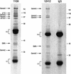
Figure 1
Figure 1
Gemin3 is in a complex with eIF2C2 in vivo. Immunoprecipitations were performed with monoclonal antibodies 11G9 or 12H12 against Gemin3 or with nonimmune mouse IgG from total HeLa cell lysates. The immunoprecipitates were resolved by SDS-PAGE and stained with Coomassie blue. The identity of immunoprecipitated proteins is shown on the left. Molecular mass markers (in kilodaltons) are shown on the right; (h.c) heavy chain of the antibody; (l.c) light chain of the antibody.
Figure 2
Figure 2
Gemin3 and Gemin4 associate with eIF2C2 in vivo and in vitro. (A) 8C7 recognizes a single band at ∼95 kD corresponding to eIF2C2, on Western blot of total HeLa cell lysate. Molecular mass markers (in kilodaltons) are shown on the right. (B) Immunoprecipitations (IP) were performed from total HeLa cell lysates with antibodies against Gemin3 (11G9), eIF2C2 (8C7), or nonimmune mouse IgG as negative control. The immunoprecipitates were analyzed by immunoblotting with monoclonal antibodies 8C7, 11G9, 17D10 (anti-Gemin4), 2B1 (anti-SMN), and 2E17 (anti-Gemin2) as indicated. Total (T) shows ∼3% of the input used for IPs. (*) Undissociated heavy and light chains of the antibodies used for IPs. (C,D) The indicated proteins were produced by in vitro transcription and translation in reticulocyte lysate, labeled with [35S]methionine, and incubated with recombinant GST–eIF2C2 (C) or GST–Gemin3, GST–Gemin4, and GST (D); bound proteins were resolved by SDS-PAGE and visualized by fluorography. Total refers to 10% of the input fraction used for binding.
Figure 2
Figure 2
Gemin3 and Gemin4 associate with eIF2C2 in vivo and in vitro. (A) 8C7 recognizes a single band at ∼95 kD corresponding to eIF2C2, on Western blot of total HeLa cell lysate. Molecular mass markers (in kilodaltons) are shown on the right. (B) Immunoprecipitations (IP) were performed from total HeLa cell lysates with antibodies against Gemin3 (11G9), eIF2C2 (8C7), or nonimmune mouse IgG as negative control. The immunoprecipitates were analyzed by immunoblotting with monoclonal antibodies 8C7, 11G9, 17D10 (anti-Gemin4), 2B1 (anti-SMN), and 2E17 (anti-Gemin2) as indicated. Total (T) shows ∼3% of the input used for IPs. (*) Undissociated heavy and light chains of the antibodies used for IPs. (C,D) The indicated proteins were produced by in vitro transcription and translation in reticulocyte lysate, labeled with [35S]methionine, and incubated with recombinant GST–eIF2C2 (C) or GST–Gemin3, GST–Gemin4, and GST (D); bound proteins were resolved by SDS-PAGE and visualized by fluorography. Total refers to 10% of the input fraction used for binding.
Figure 3
Figure 3
∼22-nt RNAs (miRNAs) are in a complex with Gemin3 and eIF2C2 in vivo. Immunoprecipitations were performed with 11G9 (anti-Gemin3), nonimmune mouse IgG, 8C7 (anti-eIF2C2), 2B1 (anti-SMN), and 12H12 (anti-Gemin3) from total HeLa cell lysates. RNA was isolated, 3′-end-labeled with [5′-32P]-pCp, and resolved by electrophoresis on 15% denaturing polyacrylamide gels. Total (T) shows RNA representing ∼3% of the input used for immunoprecipitations (IPs). The lane marked M contains 32P-labeled pBR322/MspI digest as size marker; nucleotide sizes are indicated on the left.
Figure 4
Figure 4
miRNPs sediment on sucrose gradients as ∼15S particles. Total HeLa cell lysate was sedimented on a 5%–20% sucrose gradient. Fractions were collected (indicated by numbers), resolved by SDS-PAGE, and analyzed by immunoblotting with 11G9 (anti-Gemin3), 17D10 (anti-Gemin4), and 8C7 (anti-eIF2C2). Total lane shows ∼5% of input used for gradient analysis; lane marked pellet shows ∼5% of the pellet from the gradient. (Bottom panel) fractions were pooled (indicated by brackets) and used for immunoprecipitations with 11G9. RNA was isolated, 3′-end-labeled with [5′-32P]-pCp, and resolved by electrophoresis on a 15% denaturing polyacrylamide gel. Nucleotide size marker (M) is shown on the right. S-values are shown.
Figure 5
Figure 5
Predicted secondary structure of putative miRNA precursors and organization of miRNA gene clusters. (A) 70 nt of genomic sequence upsteam and 70 nt downstream of the cloned miRNAs were used to predict the secondary structure of putative miRNA precursors, using the computer program mfold (Mathews et al. 1999). The size of the putative miRNA precursors was then reduced to ∼80 nt, and this sequence was used again for secondary structure prediction by mfold. The putative precursor of miR-91 also contains the sequence of miR-17 on the opposite strand (colored in blue). This finding is similar to the C. elegans miR-56 and miR-56*, which are processed from the same precursor, although only miR-56 is thought to be the functional miRNA (Nelson et al. 2001). Chromosomal location is indicated. Red areas represent mature miRNAs. (B) The putative miRNA precursors are represented as boxes, and the mature miRNAs identified in this study as part of miRNPs are indicated in red. Previously reported miRNAs (Lagos-Quintana et al. 2001) not found in this study are shown in blue. The chromosomal location is indicated on the right. The miRNA cluster on chromosome 13 contains two miRNAs (miR-91 and miR-92) not previously identified; miRNA-91 and miR-17 (colored in blue) are presumably derived from the same precursor. The size of clusters in nucleotides (nt) is shown.
Figure 5
Figure 5
Predicted secondary structure of putative miRNA precursors and organization of miRNA gene clusters. (A) 70 nt of genomic sequence upsteam and 70 nt downstream of the cloned miRNAs were used to predict the secondary structure of putative miRNA precursors, using the computer program mfold (Mathews et al. 1999). The size of the putative miRNA precursors was then reduced to ∼80 nt, and this sequence was used again for secondary structure prediction by mfold. The putative precursor of miR-91 also contains the sequence of miR-17 on the opposite strand (colored in blue). This finding is similar to the C. elegans miR-56 and miR-56*, which are processed from the same precursor, although only miR-56 is thought to be the functional miRNA (Nelson et al. 2001). Chromosomal location is indicated. Red areas represent mature miRNAs. (B) The putative miRNA precursors are represented as boxes, and the mature miRNAs identified in this study as part of miRNPs are indicated in red. Previously reported miRNAs (Lagos-Quintana et al. 2001) not found in this study are shown in blue. The chromosomal location is indicated on the right. The miRNA cluster on chromosome 13 contains two miRNAs (miR-91 and miR-92) not previously identified; miRNA-91 and miR-17 (colored in blue) are presumably derived from the same precursor. The size of clusters in nucleotides (nt) is shown.

References

    1. Aravin AA, Naumova NM, Tulin AV, Vagin VV, Rozovsky YM, Gvozdev VA. Double-stranded RNA-mediated silencing of genomic tandem repeats and transposable elements in the D. melanogaster germline. Curr Biol. 2001;11:1017–1027. - PubMed
    1. Bass BL. Double-stranded RNA as a template for gene silencing. Cell. 2000;101:235–238. - PubMed
    1. Bernstein E, Caudy AA, Hammond SM, Hannon GJ. Role for a bidentate ribonuclease in the initiation step of RNA interference. Nature. 2001;409:363–366. - "VSports注册入口" PubMed
    1. Catalanotto C, Azzalin G, Macino G, Cogoni C. Gene silencing in worms and fungi. Nature. 2000;404:245. - "V体育官网" PubMed
    1. Charroux B, Pellizzoni L, Perkinson RA, Shevchenko A, Mann M, Dreyfuss G. Gemin3: A novel DEAD box protein that interacts with SMN, the spinal muscular atrophy gene product, and is a component of gems. J Cell Biol. 1999;147:1181–1194. - PMC - PubMed

"VSports app下载" Publication types

MeSH terms

Substances