Skip to main page content
U.S. flag

An official website of the United States government

Dot gov

The . gov means it’s official. Federal government websites often end in . gov or . mil VSports app下载. Before sharing sensitive information, make sure you’re on a federal government site. .

Https

The site is secure. The https:// ensures that you are connecting to the official website and that any information you provide is encrypted and transmitted securely. V体育官网.

. 2001 Dec;109(2):195-204.
doi: 10.1016/s0925-4773(01)00524-x.

SNT-1/FRS2alpha physically interacts with Laloo and mediates mesoderm induction by fibroblast growth factor

Affiliations

SNT-1/FRS2alpha physically interacts with Laloo and mediates mesoderm induction by fibroblast growth factor

VSports手机版 - J Hama et al. Mech Dev. 2001 Dec.

Abstract (VSports app下载)

Members of the fibroblast growth factor (FGF) ligand family play a critical role in mesoderm formation in the frog Xenopus laevis. While many components of the signaling cascade triggered by FGF receptor activation have been identified, links between these intracellular factors and the receptor itself have been difficult to establish. We report here the characterization of Xenopus SNT-1 (FRS2alpha), a scaffolding protein previously identified as a mediator of FGF activity in other biological contexts. SNT-1 is widely expressed during early Xenopus development, consistent with a role for this protein in mesoderm formation. Ectopic SNT-1 induces mesoderm in Xenopus ectodermal explants, synergizes with low levels of FGF, and is blocked by inhibition of Ras activity, suggesting that SNT-1 functions to transmit signals from the FGF receptor during mesoderm formation. Furthermore, dominant-inhibitory SNT-1 mutants inhibit mesoderm induction by FGF, suggesting that SNT-1 is required for this process VSports手机版. Expression of dominant-negative SNT-1 in intact embryos blocks mesoderm formation and dramatically disrupts trunk and tail development, indicating a requirement for SNT-1, or a related factor inhibited by the mutant construct, during axis formation in vivo. Finally, we demonstrate that SNT-1 physically associates with the Src-like kinase Laloo, and that SNT-1 activity is required for mesoderm induction by Laloo, suggesting that SNT-1 and Laloo function as components of a signaling complex during mesoderm formation in the vertebrate. .


Figures
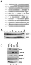
Fig. 1
Fig. 1
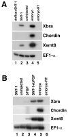
Sequence and expression of Xenopus SNT-1. (A) Alignment of the putative human and Xenopus SNT-1 amino acid sequences. Identical amino acids are shown in filled boxes. The conserved phosphotyrosine binding (PTB) domains are indicated. The location of the six tyrosine residues mutated in 6YF are highlighted (see below) (B) RT-PCR analysis of XSNT-1 expression during development. (C) RT-PCR analysis of XSNT-1 expression in gastrula stage explants (stage 10.5). The “-RT” lane contains all reagents except reverse transcriptase and was used as a negative control. Ornithine decarboxylase (ODC) is used as a loading control (Bassez et al., 1990). Xbrachyury (Xbra) is a panmesodermal marker at this stage (Smith et al., 1991); Chordin is a dorsal mesodermal marker (Sasai et al., 1994); Xwnt8 is a ventrolateral mesodermal marker (Smith and Harland, 1991; Christian et al., 1991).
Fig. 2
Fig. 2
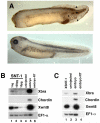
Effects of SNT-1 misexpression. (A) top: Lateral view of stage 35 embryo injected with 1ng human SNT-1 RNA in the animal pole. bottom: lateral view of uninjected sibling embryo. Anterior is to left. SNT-1 injection results in a loss of anterior structures, including eyes, the generation of small, ectopic tail-like structures (arrowheads), and occasional enlargement of the proctodeum. (B) Human SNT-1 induces mesoderm in animal caps. RT-PCR analysis of animal caps dissected at late blastula stages and cultured until midgastrula stages. Animal caps were isolated from embryos injected with SNT-1 RNA at the 2-cell stage, as listed. (C) Xenopus SNT-1 (XSNT-1) induces mesoderm in animal caps. RT-PCR analysis of animal caps dissected at late blastula stages and cultured until midgastrula stages. Animal caps were isolated from embryos injected at the 2-cell stage with 1 ng XSNT-1.
Fig. 3
Fig. 3
SNT-1 functions within a signaling cascade that includes FGF and Ras. (A) SNT-1 activity is inhibited by expression of a dominant inhibitory Ras construct. 1 ng SNT-1 and 1ng dominant inhibitory Ras (dnRas) RNA were injected, as listed. (B) SNT-1 and basic FGF (bFGF) protein synergize in the mesoderm induction assay. 50 pg SNT-1 and 1 ng/mL bFGF were used, as listed. RT-PCR analysis of animal caps cultured until midgastrula stages.
Fig. 4
Fig. 4
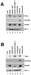
SNT-1 mutants function as putative dominant-negative reagents. (A) Inhibition of FGF-mediated mesoderm induction by ΔMT, and subsequent rescue with full-length SNT-1. (B) Inhibition of FGF-mediated mesoderm induction by 6YF, and subsequent rescue with full-length SNT-1. RT-PCR analysis of animal caps cultured until midgastrula stages. 2 ng ΔMT, 1 ng 6YF, and 1 ng SNT-1 RNA were injected, as listed. 25 ng/mL bFGF was added, as listed.
Fig. 5
Fig. 5
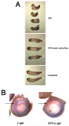
The SNT-1 mutant 6YF inhibits mesoderm induction and normal development in the Xenopus embryo. (A) Stage 32 embryos, injected with 500 pg 6YF RNA alone (top panel), co-injected with 500 pg 6YF and 40 pg activated Ras (const. active Ras) RNA, or uninjected. Anterior is to left. Bottom two embryos in top panel are dorsal views, showing the open blastopore remnant; all other views are lateral. (B) β-gal staining and whole-mount in situ hybridization of midgastrula stage albino embryos using an antisense Xbrachyury probe; vegetal-posterior view. The embryo on the left was injected with 100pg β-galactosidase RNA in the equatorial region of one blastomere at the 2-cell stage; note overlap between Xbrachyury expression (blue) and the red β-gal product (arrow). The embryo on the right was injected with both 100pg β-galactosidase RNA and 500 pg 6YF RNA in the same region; note the presence of β-gal product in the gap in Xbrachyury expression (arrow).
Fig. 6
Fig. 6
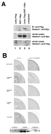
Physical association between SNT-1 and Laloo. (A) Western blot analysis of SNT-1/Laloo co-immunoprecipitation. (B) Yeast 2-hybrid analysis demonstrating interaction between the SH4/SH3 domains of Laloo and SNT-1. Expression vectors for Myc-tagged Gal4 DNA binding domain fusion proteins (Laloo or FGFR1 segments) and Gal4 activation domain fusion proteins (SNT-1 or PLCγ-SH2 domain) were cotransformed in yeast and plated onto synthetic medium without leucine/tryptophan to measure cotransformation efficiency or onto medium without leucine/tryptophan/histidine + 25mM 3-amino-1,2,4-triazole (AT) to select clones displaying fusion protein interaction. No proteins tested interacted with the negative control PLCγ fusion protein (not shown). Bottom panel: LalooSH4/3, LalooSH4, and LalooSH3-30 protein are expressed in yeast. Yeast protein extracts were immunoprecipitated with anti-Myc monoclonal antibody and analyzed by Western blot with a monoclonal antibody against the GAL4 DNA binding domain.
Fig. 7
Fig. 7
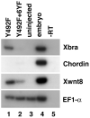
Expression of dominant-negative SNT-1 inhibits mesoderm induction by the activated Laloo mutant Y492F. RT-PCR analysis of animal caps cultured until midgastrula stages. 100 pg Y492F RNA and 1 ng 6YF RNA were injected, as listed.
Fig. 8
Fig. 8
Model depicting the role of SNT-1 and Laloo in FGF signaling during early vertebrate development. Arrows do not necessarily indicate direct molecular interactions. See text for details.

References

    1. Amaya E, Kroll KL. Transgenic Xenopus embryos from sperm nuclear transplantations reveal FGF signaling requirements during gastrulation. Development. 1996;122:3173–3183. - PubMed
    1. Amaya E, Musci TJ, Kirschner MW. Expression of a dominant negative mutant of the FGF receptor disrupts mesoderm formation in Xenopus embryos. Cell. 1991;66:257–270. - VSports在线直播 - PubMed
    1. Bassez T, Paris J, Omilli F, Dorel C, Osborne HB. Post-transcriptional regulation of ornithine decarboxylase in Xenopus laevis oocytes. Development. 1990;110:955–962. - "VSports手机版" PubMed
    1. Brewster R, Mullor JL, Ruiz I Altaba A. Gli2 functions in FGF signaling during antero-posterior patterning. Development. 2000;127:4395–4405. - PubMed
    1. Carballada R, Yasuo H, Lemaire P. Phosphatidylinositol-3 kinase acts in parallel to the ERK MAP kinase in the FGF pathway during Xenopus mesoderm induction. Development. 2001;128:35–44. - PubMed

"VSports注册入口" Publication types

MeSH terms

LinkOut - more resources