Skip to main page content
U.S. flag

An official website of the United States government

Dot gov

The . gov means it’s official. Federal government websites often end in . gov or . mil. Before sharing sensitive information, make sure you’re on a federal government site VSports app下载. .

Https

The site is secure V体育官网. The https:// ensures that you are connecting to the official website and that any information you provide is encrypted and transmitted securely. .

. 2001 Dec;69(12):7652-62.
doi: 10.1128/IAI.69.12.7652-7662.2001.

Arginine-143 of Yersinia enterocolitica YopP crucially determines isotype-related NF-kappaB suppression and apoptosis induction in macrophages

Affiliations

VSports注册入口 - Arginine-143 of Yersinia enterocolitica YopP crucially determines isotype-related NF-kappaB suppression and apoptosis induction in macrophages

K Ruckdeschel et al. Infect Immun. 2001 Dec.

Abstract

Pathogenic Yersinia spp. counteract host defense mechanisms by modulating the cellular signal relay in response to infection. Subversion of the antiapoptotic NF-kappaB signaling pathway by the Yersinia enterocolitica virulence protein YopP crucially determines the induction of apoptosis in Yersinia-infected macrophages. Here, we analyzed a panel of pathogenic, phylogenetically distinct Y. enterocolitica serotypes for their abilities to trigger macrophage apoptosis. Y. enterocolitica from the highly pathogenic serogroup O8 was substantially more effective in apoptosis induction than Yersinia from the serogroups O3 and O9. Complementation of yopP-knockout mutants revealed that this effect was specifically conferred by the serogroup O8 YopP. The amino acid sequences of YopPO8 and YopPO9 share 94% identity, and both YopP isotypes were found to interact with the NF-kappaB-activating kinase IKKbeta in macrophages. However, selectively, YopPO8 mediated efficient inhibition of IKKbeta activities, which led to substantial suppression of NF-kappaB activation. To localize the YopPO8-related effector domain, we interchanged stretches of amino acids and single amino acid residues between YopPO8 and YopPO9 VSports手机版. Functional characterization of the resulting mutants revealed a major role of the arginine-143 residue in determining the inhibitory impact of YopP on IKKbeta activity and survival of macrophages. .


Figures
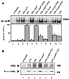
FIG. 1
FIG. 1
YopPO8 efficiently triggers macrophage apoptosis and interferes with NF-κB activation. (A) NF-κB suppression and apoptosis induction. J774A.1 cells were left untreated (lane 1) or were stimulated with LPS (lane 2), virulence plasmid-cured WA-ΔpYV (lane 3), wild-type WA from serogroup O8 (lane 4), yopPO8-negative WA-ΔyopP (lane 5), YopPO8-producing WA-ΔyopP/+PO8 (lane 6), YopPO9-producing WA-ΔyopP/+PO9 (lane 7), WA-ΔΣyop producing no effector Yops (lane 8), WA-ΔΣyop/+PO8 producing only YopPO8 (lane 9), WA-ΔΣyop/+PO9 producing only YopPO9 (lane 10), or wild-type E40 from serogroup O9 (lane 11). The NF-κB activities were determined by EMSA 90 min after infection (upper panel). Only sections of the autoradiogram containing the NF-κB–DNA complexes are shown. The EMSA shows data from one experiment representative of five performed. Apoptosis was assayed 6 h after onset of infection by staining cells with annexin V and counting apoptotic cells by fluorescence microscopy (lower panel). Results are expressed as mean percentages ± standard deviations from three independent experiments. (B) IKKβ activity assay. Cell extracts from 1.25 × 10⁷ untreated cells (lane 1) or cells treated with LPS (lane 2), wild-type WA from serogroup O8 (lane 3), yopPO8-negative WA-ΔyopP (lane 4), YopPO8-producing WA-ΔyopP/+PO8 (lane 5), or YopPO9-producing WA-ΔyopP/+PO9 (lane 6) for 30 min were incubated with anti-IKKβ antibodies to precipitate IKKβ. IKKβ activities were assayed by measuring the abilities of the immunocomplexes to radioactively phosphorylate recombinant GST-IκBα. Kinase reaction samples were subjected to SDS-PAGE and transferred to PVDF membrane. The upper part of the membrane was immunoblotted with anti-IKK antibodies (upper panel). The double band appearing in lanes 2 to 6 may reflect phosphorylated and nonphosphorylated forms of IKKβ. The lower part of the membrane including GST-ΙκBα was analyzed by autoradiography (lower panel). The results shown are from one representative experiment out of three performed. WB, Western blot; KA, kinase assay.
FIG. 2
FIG. 2
Alignment of the deduced amino acid sequences from YopPO8 (GenBank accession no. AF336309) and YopPO9 (GenBank accession no. AF102990). Amino acid differences are marked in boldface. The corresponding amino acids of the internal yopP BamHI restriction sites are underlined. Dark shading denotes the proposed isotype-related YopP effector site (arginine-143, YopPO8; serine-143, YopPO9). Light shading indicates the amino acid residues of the proposed cysteine protease-related catalytic triad (histidine-109, glutamic acid-128, and cysteine-172).
FIG. 3
FIG. 3
Interchange of amino acids 44 to 214 reverses YopPO8 and YopPO9 isotype-dependent effector functions. J774A.1 cells were left untreated (lane 1) or were infected with yopPO9-negative E40-ΔyopP (lane 2), YopPO8-producing E40-ΔyopP/+PO8 (lane 3), YopPO9-producing E40-ΔyopP/+PO9 (lane 4), E40-ΔyopP/+PO8(44–214PO9) producing a YopPO8 construct in which the internal amino acids 44 to 214 were substituted for by the respective amino acids from YopPO9 (lane 5), and E40-ΔyopP/+PO9(44–214PO8) producing a YopPO9 construct in which internal amino acids 44 to 214 were substituted for by the respective amino acids from YopPO8 (lane 6). The NF-κB activities were determined 90 min after infection by EMSA (upper panel). Only sections of the autoradiogram containing the NF-κB–DNA complexes are shown. The EMSA shows data from one experiment representative of three performed. Apoptosis was assayed 6 h after onset of infection by staining cells with annexin V and counting apoptotic cells by fluorescence microscopy (lower panel). Results are expressed as mean percentages ± standard deviations from three independent experiments.
FIG. 4
FIG. 4
Mapping of the YopPO8 isotype-related effector domain by interchange of amino acids between YopPO9 and YopPO8. Mutagenesis of YopPO9 (lane 1) was accomplished by replacement of an internal 514-bp yopPO9 DNA fragment by diverse yopPO9/yopPO8 hybrid sequences for this region (lanes 2 to 8). The DNA region encodes amino acids 44 to 214 of YopP. The fragments were generated by PCR and introduced into the internal yopPO9 BamHI restriction sites, which encompass the 514-bp region. In lanes 2 to 8, only the amino acid sequences introduced from YopPO8, but not the endogenous YopPO9 amino acid sequences, are displayed. Mutagenesis of YopPO8 (lane 9) was conducted by the same method, resulting in substitution of multiple or single amino acids of YopPO8 by residues from YopPO9 (lanes 10 to 13). Apoptosis was assayed 6 h after onset of infection by staining cells with annexin V and analyzing apoptotic cells by fluorescence microscopy [apoptosis: (+), 20 to 40%; +, 40 to 60%; ++, 60 to 80%; +++, >80%].
FIG. 5
FIG. 5
Arginine-143 critically determines YopP effector functions. (A) NF-κB suppression and apoptosis induction. J774A.1 cells were infected with YopPO9-producing E40-ΔyopP/+PO9 (lane 1), YopPO8-producing E40-ΔyopP/+PO8 (lane 2), E40-ΔyopP/+PO8(R143S) producing a YopPO8 construct in which arginine-143 was substituted for by serine (lane 3), or E40-ΔyopP/+PO9(S143R) producing a YopPO9 construct in which serine-143 was substituted for by arginine (lane 4). The NF-κB activities were determined 90 min after infection by EMSA (upper panel). Only sections of the autoradiogram containing the NF-κB–DNA complexes are shown. The EMSA shows data from one experiment representative of three performed. Apoptosis was assayed 6 h after onset of infection by staining cells with annexin V and counting apoptotic cells by fluorescence microscopy (lower panel). Results are expressed as mean percentages ± standard deviations from three independent experiments. (B) IKKβ activity assay. Cell extracts from 108 cells infected with YopPO8-producing E40-ΔyopP/+PO8 (lane 1), YopPO9-producing E40-ΔyopP/+PO9 (lane 2), YopPO9(S143R)-producing E40-ΔyopP/+PO9(S143R) (lane3), or YopPO8(R143S)-producing E40-ΔyopP/+PO8(R143S) (lane 4) for 30 min were incubated with anti-IKKβ antibodies to precipitate IKKβ. IKKβ activities were assayed by measuring the abilities of the immunocomplexes to radioactively phosphorylate recombinant GST-IκBα. Kinase reaction samples were subjected to SDS-PAGE and transferred to PVDF membrane. The upper part of the membrane was immunoblotted with anti-IKK antibodies (upper panel). The lower part of the membrane including GST-ΙκBα was analyzed by autoradiography (lower panel). The results shown are from one representative experiment out of three performed. WB, Western blot; KA, kinase assay.
FIG. 6
FIG. 6
Targeting of macrophage IKKβ by the diverse YopP constructs. (A.) Interaction of YopP with macrophage IKKβ. J774A.1 cells were infected with yopPO9-negative E40-ΔyopP (lane 1), YopPO8-producing E40-ΔyopP/+PO8 (lane 2), YopPO9-producing E40-ΔyopP/+PO9 (lane 3), WA-ΔΣyop producing no effector Yops (lane 4), WA-ΔΣyop/+PO8 producing only YopPO8 (lane 5), WA-ΔΣyop/+PO9 producing only YopPO9 (lane 6), WA-ΔΣyop/+PO8(R143S) producing only the YopPO8 construct with the mutation of arginine-143 to serine (lane 7), or WA-ΔΣyop/+PO9(S143R) producing only the YopPO9 construct with the mutation of serine-143 to arginine (lane 8). After 60 min, cells were lysed, and YopP was immunoprecipitated with polyclonal anti-YopE antibody recognizing the YopE138-YopP fusion proteins. Immunocomplexes were subjected to SDS-PAGE and transferred to PVDF membrane. One part of the membrane was immunoblotted with anti-YopE antibodies, recognizing the YopP fusion proteins (lower panel), and the other part was immunoblotted with anti-ΙKK antibodies (upper panel). (B) Quantification of amounts of cytoplasmic YopP. J774A.1 cells were infected with WA-ΔΣyop producing no effector Yops (lane 1), WA-ΔΣyop/+PO8 producing only YopPO8 (lane 2), or WA-ΔΣyop/+PO8(R143S) producing only the YopPO8 construct with the mutation of arginine-143 to serine (lane 3). After 60 min, cells were lysed under conditions that selectively lyse the cells. The lysates were subjected to SDS-PAGE and transferred to PVDF membrane. One part of the membrane was immunoblotted with anti-YopE antibodies, recognizing the YopP fusion proteins (upper panel), and the other part was immunoblotted with control anti-MEK1 antibodies to confirm equal loading with cellular lysates (lower panel). Nonsaturated immunoreactive bands were visualized with enhanced chemiluminescence reagents. The optical densities (OD) of the YopP bands were quantified, and the values obtained are indicated. The results shown are from one representative experiment out of three performed.

References

    1. Aliprantis A O, Yang R B, Weiss D S, Godowski P, Zychlinsky A. The apoptotic signaling pathway activated by Toll-like receptor-2. EMBO J. 2000;19:3325–3336. - PMC - PubMed
    1. Baichwal V R, Baeuerle P A. Apoptosis: activate NF-κB or die? Curr Biol. 1997;7:R94–R96. - PubMed (V体育ios版)
    1. Baldwin A S., Jr The transcription factor NF-κB and human disease. J Clin Investig. 2001;107:3–6. - PMC - PubMed
    1. Bliska J B. Yop effectors of Yersinia spp. and actin rearrangements. Trends Microbiol. 2000;8:205–208. - PubMed
    1. Boland A, Cornelis G R. Role of YopP in suppression of tumor necrosis factor alpha release by macrophages during Yersinia infection. Infect Immun. 1998;66:1878–1884. - PMC - PubMed

Publication types

V体育ios版 - MeSH terms