Skip to main page content
U.S. flag

An official website of the United States government

Dot gov

The . gov means it’s official. Federal government websites often end in VSports app下载. gov or . mil. Before sharing sensitive information, make sure you’re on a federal government site. .

Https

The site is secure. The https:// ensures that you are connecting to the official website and that any information you provide is encrypted and transmitted securely. V体育官网.

. 2001 Nov;12(11):3690-702.
doi: 10.1091/mbc.12.11.3690.

Autophagosome requires specific early Sec proteins for its formation and NSF/SNARE for vacuolar fusion (VSports注册入口)

Affiliations
Free PMC article

Autophagosome requires specific early Sec proteins for its formation and NSF/SNARE for vacuolar fusion

N Ishihara et al. Mol Biol Cell. 2001 Nov.
Free PMC article

Abstract

Double membrane structure, autophagosome, is formed de novo in the process of autophagy in the yeast Saccharomyces cerevisiae, and many Apg proteins participate in this process. To further understand autophagy, we analyzed the involvement of factors engaged in the secretory pathway VSports手机版. First, we showed that Sec18p (N-ethylmaleimide-sensitive fusion protein, NSF) and Vti1p (soluble N-ethylmaleimide-sensitive fusion protein attachment protein, SNARE), and soluble N-ethylmaleimide-sensitive fusion protein receptor are required for fusion of the autophagosome to the vacuole but are not involved in autophagosome formation. Second, Sec12p was shown to be essential for autophagy but not for the cytoplasm to vacuole-targeting (Cvt) (pathway, which shares mostly the same machinery with autophagy. Subcellular fractionation and electron microscopic analyses showed that Cvt vesicles, but not autophagosomes, can be formed in sec12 cells. Three other coatmer protein (COPII) mutants, sec16, sec23, and sec24, were also defective in autophagy. The blockage of autophagy in these mutants was not dependent on transport from endoplasmic reticulum-to-Golgi, because mutations in two other COPII genes, SEC13 and SEC31, did not affect autophagy. These results demonstrate the requirement for subgroup of COPII proteins in autophagy. This evidence demonstrating the involvement of Sec proteins in the mechanism of autophagosome formation is crucial for understanding membrane flow during the process. .


Figures
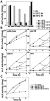
Figure 1
Figure 1
Sec18p and Vti1p are essential for the autophagy. (A) Cells expressing Pho8Δ60p (wild-type [wt], sec18, sec17, vti1-11, and cdc48-3) cultured in YPD at 23°C (open bar) were shifted to SD(-N) and cultured for 4 h at 23°C (shaded bar) or 34°C (filled bar). The ALP activity of the lysate was measured to estimate autophagic activity. (B) Time course of autophagy. Wild-type, sec18, sec17, and vti1-11 cultured in YPD at 23°C were shifted to SD(-N) at 23°C (circle), 34°C (square), or 37°C (triangle). Cells cultured in SD(-N) for 3 h were shifted from 23 to 34°C (open circle), from 34 to 23°C (open square), or from 37 to 23°C (open triangle). (C) Recovery of autophagy in sec18. Wild-type (wt) and sec18 cells cultured in YPD at 23°C were shifted to SD(-N) at 23°C (wt, open circle; sec18, filled circle) or 30°C (sec18, filled square). sec18 cells cultured in SD(-N) at 30°C for 2 h were shifted to 23°C (open square).
Figure 2
Figure 2
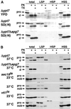
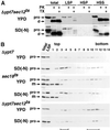
Accumulation of autophagosomes in sec18 and vti1 mutants. (A) Fractionation of autophagosomes and Cvt vesicles. Δypt7 cells growing in YPD or Δypt7 cells and Δypt7Δapg7 cells starved in SD(-N) were converted to spheroplasts and lysed. Cleared lysates were obtained by centrifugation at 500 × g for 5 min and were then centrifuged at 13,000 × g for 15 min to generate the LSP. The supernatants were further centrifuged at 100,000 × g for 1 h to separate the HSP and HSS. Each fraction was treated with 100 μg/ml proteinase K (PK) with or without 1% Triton X-100 (TX) and then analyzed by immunoblotting with antiserum to API. (B) Biochemical detection of autophagosomes in sec18 and vti1-11. Δypt7 and Δypt7Δapg7 cells were cultured at 37°C, and sec18 and vti1-11 were cultured at 23 or 37°C in SD(-N) for 3 h. Cell lysis under milder conditions, subcellular fractionation, and protease treatment were performed as described in MATERIALS AND METHODS. Each sample was subjected to SDS-PAGE and immunoblotting with anti-API serum. Precursor (pro) and mature forms (m) of API are indicated. d, the proteinase K digestion products of proAPI.
Figure 3
Figure 3
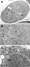
Electron micrographs of autophagosomes in the sec18 mutant. Wild-type (A) and sec18 (B and C) cells were cultured in SD(-N) containing PMSF at 37°C for 2.5 h and fixed by the freeze substitution technique as described in MATERIALS AND METHODS. Higher magnification images of the autophagosomes in sec18 are shown in D and E. Arrows, autophagosome; arrowheads, autophagic body; V, vacuole. Bars, 500 nm (A–C); 100 nm (D and E).
Figure 4
Figure 4
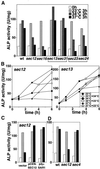
Autophagic activity in the COPII mutants. (A) Cells expressing Pho8Δ60p (wild-type [wt], sec12, sec16, sec13, sec31, sec23, or sec24) were cultured in YPD at 23°C (open bar), transferred to SD(-N) at 23°C (shaded bar) or 34°C (gray bar) or 37°C for 3 h (filled bar), lysed, and assessed for ALP activity. (B) Time course of autophagy in sec12 and sec13. sec12 and sec13 cells grown in YPD at 23°C were cultured in SD(-N) at 23°C (circle), 34°C (square), or 37°C (triangle). The cells cultured in SD(-N) for 3 h were shifted from 23 to 34°C (open circle), from 34 to 23°C (open square), and from 37 to 23°C (open triangle). (C) sec12 cells carrying pRS316 (vector), pSHY6-4 (pCEN-SEC12), or pANY2-7 (p2 μ-SAR1) grown in SC(-Ura) at 23°C were cultured in YPD for 4 h at 23°C (open bar) and then transferred to SD(-N) for 4 h at 23°C (shaded bar) or 34°C (filled bar). (D) Wild-type (wt), sec12, or sec4 cells grown in YPD at 23°C (open bar) were cultured in SD(-N) at 23°C (shaded bar) or 34°C (filled bar) for 2 h. The ALP activities of the lysate were measured as described in MATERIALS AND METHODS.
Figure 5
Figure 5
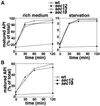
API transport in early secretory mutants. (A) Wild-type (wt, open circle), sec12 (filled circle), sec13 (open square), or sec18 (filled square) cultured to the midlog phase in SC(-Met) at 23°C were shifted to 37°C for 10 min, labeled with [35S]methionine for 20 min at 37°C, and then chased after addition of SC(-Met) (rich medium) or SD(-N) (starvation) containing methionine/cysteine at 37°C for the times indicated. Immunoprecipitation with anti-API serum was carried out as described in MATERIALS AND METHODS and analyzed by SDS-PAGE and a Bioimage analyzer, BAS2000. (B) Wild-type, sec12, and sec18 cells cultured in SC(-Met) at 23°C were heated to 37°C for 1 h, labeled for 20 min at that temperature, and then chased in a rich medium for the time indicated. Immunoprecipitation was carried out as described in MATERIALS AND METHODS.
Figure 6
Figure 6
API transport was impaired in sec12Δcvt9 cells. (A) Wild-type (wt), Δcvt9, sec12Δcvt9, and sec12 cells grown in YPD at 23°C were cultured at 37°C in YPD or SD(-N) for 3 h, lysed, and analyzed by immunoblotting with anti-API serum. (B) The mutant cells in A were cultured in YPD at 23°C (open bar), transferred to SD(-N) at 23°C (shaded bar) or 34°C (filled bar) for 3 h, lysed, and assessed for ALP activity.
Figure 7
Figure 7
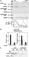
Fractionation of autophagosomes and Cvt vesicles by density gradient centrifugation. (A) Δypt7, Δypt7Δapg7, and Δypt7Δcvt9 cells were cultured in YPD or SD(-N) at 30°C. The LSP fractions were prepared as described in Figure 2 and were loaded on top of a 10–50% OptiPrep gradient and centrifuged for 16 h at 174,000 × g. Fourteen fractions were collected from the top of the tubes. An equal volume of each fraction was subjected to immunoblotting with anti-API serum. As a control, one-sixth of the cell lysate (total) and half of the LSP were used. Organelle markers of the vacuole (proteinase B, PrB), endosomes (Pep12p), and mitochondria (F1β) were detected by immunoblotting of starved Δypt7 cells and estimated by densitometry. (B) The relative amounts of proAPI and Pho8Δ60p in fractions 3 and 4 or fractions 9–11 in the density gradients in A were determined by immunoblotting with their respective antisera. (C) Fractions 3 and 4 from starved cells (AP) or fractions 9–11 from YPD-grown cells (Cvt) in A were pooled, treated with proteinase K (PK) with or without Triton X-100 (TX), and then subjected to immunoblotting with anti-API or anti-Bmh1p antiserum.
Figure 8
Figure 8
Autophagosome formation in sec12 and sec18 cells. (A) Δypt7 sec12 cells were cultured in YPD or SD(-N) at 37°C for 3 h. Fractionation and protease treatment were performed as in Figure 2. (B) Δypt7, sec18, and Δypt7 sec12 cells precultured in YPD at 23°C were cultured in YPD or SD(-N) at 37°C for 3 h. Density gradient centrifugation and immunoblotting were carried out as described in Figure 7. As a control, one-tenth of the total cell lysate and half of the LSP were also analyzed.
Figure 9
Figure 9
Electron micrographs of sec12 under starvation conditions. sec12 mutant cells were cultured in SD(-N) containing PMSF at 37°C for 2.5 h and prepared for electron microscopy as described in MATERIALS AND METHODS. Higher magnification images are shown in B and C. Arrows, Cvt vesicle; arrowheads, Cvt body; V, vacuole. Bars, 500 nm (A); 100 nm (B and C).

"VSports最新版本" References

    1. Abeliovich H, Darsow T, Emr SD. Cytoplasm to vacuole trafficking of aminopeptidase I requires a t-SNARE-Sec1p complex composed of Tlg2p and Vps45p. EMBO J. 1999;18:6005–6016. - PMC - PubMed
    1. Adams A, Gottschling DE, Kaiser CA, Stearns T. Methods in Yeast Genetics. Cold Spring Harbor, NY: Cold Spring Harbor Laboratory Press; 1997.
    1. Baba M, Osumi M, Scot SV, Klionsky DJ, Ohsumi Y. Two distinct pathways for targeting proteins from the cytoplasm to the vacuole/lysosome. J Cell Biol. 1997;139:1687–1695. - PMC - PubMed
    1. Baba M, Takeshige K, Baba N, Ohsumi Y. Ultrastructural analysis of the autophagic process in yeast: detection of autophagosomes and their characterization. J Cell Biol. 1994;124:903–913. - PMC - PubMed
    1. Barlowe C, Orc L, Yeung T, Hosobuchi M, Hamamoto S, Salama N, Rexach MF, Ravazzola M, Amherdt M, Scheckman R. COPII: a membrane coat formed by Sec proteins that drive vesicle budding from the endoplasmic reticulum. Cell. 1994;77:895–907. - PubMed (V体育官网)

Publication types

MeSH terms

VSports手机版 - Substances