<tt lang="P3nWn"></tt> Skip to main page content
U.S. flag

An official website of the United States government

Dot gov

The . gov means it’s official. Federal government websites often end in . gov or . mil VSports app下载. Before sharing sensitive information, make sure you’re on a federal government site. .

Https

The site is secure. The https:// ensures that you are connecting to the official website and that any information you provide is encrypted and transmitted securely. V体育官网.

. 2001 Oct;13(10):2211-24.
doi: 10.1105/tpc.010085.

The disease resistance signaling components EDS1 and PAD4 are essential regulators of the cell death pathway controlled by LSD1 in Arabidopsis (V体育2025版)

Affiliations

The disease resistance signaling components EDS1 and PAD4 are essential regulators of the cell death pathway controlled by LSD1 in Arabidopsis

C Rustérucci et al. Plant Cell. 2001 Oct.

Abstract (V体育官网入口)

Specific recognition of pathogens is mediated by plant disease resistance (R) genes and translated into a successful defense response. The extent of associated hypersensitive cell death varies from none to an area encompassing cells surrounding an infection site, depending on the R gene activated. We constructed double mutants in Arabidopsis between positive regulators of R function and a negative regulator of cell death, LSD1, to address whether genes required for normal R function also regulate the runaway cell death observed in lsd1 mutants. We report here that EDS1 and PAD4, two signaling genes that mediate some but not all R responses, also are required for runaway cell death in the lsd1 mutant. Importantly, this novel function of EDS1 and PAD4 is operative when runaway cell death in lsd1 is initiated through an R gene that does not require EDS1 or PAD4 for disease resistance. NDR1, another component of R signaling, also contributes to the control of plant cell death. The roles of EDS1 and PAD4 in regulating lsd1 runaway cell death are related to the interpretation of reactive oxygen intermediate-derived signals at infection sites. We further demonstrate that the fate of superoxide at infection sites is different from that observed at the leading margins of runaway cell death lesions in lsd1 mutants. VSports手机版.


Figures
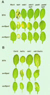
Figure 1.
Figure 1.
Lesion Phenotypes of Plant Lines after BTH Treatment or Bacterial Pathogen Inoculation. Leaves of 4-week-old wild-type, single mutant, or double mutant plants were sprayed with 0.35 mM BTH or infiltrated on one side with low titer suspensions (105 colony-forming units/mL) of P. syringae pv DC3000 expressing avrRps4 or avrRpm1. See Methods for further details. Leaves were photographed after 6 days of incubation, and each leaf is representative of 12 to 15 leaves. All treatments were repeated with similar results. (A) Phenotypes of plant lines in accession Ws-0. (B) Phenotypes of plant lines in accession Col-0.
Figure 2.
Figure 2.
Bacterial Growth in Wild-Type, Single Mutant, and Double Mutant Plants. Growth of P. syringae pv DC3000 expressing avrRps4 or avrRpm1 extracted from leaves at 0 (open bars) and 3 (closed bars) days after inoculation (initial titer, 105 colony-forming units/mL). Data from Ws-0
Figure 3.
Figure 3.
Infection Phenotypes of Plant Cotyledons Inoculated with Pp Isolates Noco2 and Emoy2. (A) Cotyledons of 10-day-old seedlings were inoculated with Noco2 or Emoy2 (5 × 10⁴ spores/mL) and stained with lactophenol–trypan blue at 6 DPI to reveal Pp mycelium and dead plant cells. (B) Trypan blue–stained cotyledons were harvested at 6 DPI, and individual plant–pathogen interaction sites were categorized as HR, trailing necrosis, or free mycelium. The percentage of each interaction type was scored from 40 to 80 cotyledons per experiment. Graphs represent the mean and ±se from three independent experiments.
Figure 4.
Figure 4.
Disease Resistance and Runaway Plant Cell Death Phenotypes in Adult Leaves after Pp Inoculation. Leaves of 4-week-old plants from wild-type, single mutant, and double mutant Ws-0 ([A] and [C]) and Col-0 ([B] and [D]) lines were inoculated by placing a 10-μL droplet of Pp spores on the top half of each leaf. Pp isolates Noco2 and Emoy2 (or Emwa1) were used for Ws-0 ([A] and [C]) and Col-0 ([B] and [D]) accession lines, respectively. Macroscopic phenotypes and corresponding trypan blue (TB) staining of plant–pathogen interaction sites are shown for whole leaves ([A] and [B]) and at ×200 magnification ([C] and [D], bottom row) at 6 DPI. Hypersensitive plant cell death in trypan blue–stained leaves is marked with black arrows. Accumulation of H2O2 at interaction sites 32 hr after inoculation of leaves in the dark was measured using DAB and is shown at ×200 magnification ([C] and [D], top row). The inset in the upper left corner of the eds1 image at (C) shows an enlarged view of DAB staining restricted to the pathogen penetration site. Images are representative of four independent experiments using at least five leaves per genotype per experiment.
Figure 5.
Figure 5.
Localized H2O2 Accumulation in Wild-Type and lsd1 Leaves after RB Application or Pp Infection. (A) A single 5-μL spot of 20 mM RB was applied to leaves of 4-week-old plants. H2O2 accumulation was measured over 7 days by staining with DAB. Leaves also were stained with lactophenol–trypan blue (TB) to measure the extent of plant cell death. Localized RB application gives rise to local H2O2 accumulation associated with a discrete patch of dead plant cells. In Ws-0, expansion of plant cell necrosis ceases by 27 hr, whereas in lsd1, the lesions expand. DAB-staining material is detected in the area of RB application but is not associated with the spreading RCD in lsd1 (black arrows). (B) Leaves of 4-week-old plants were inoculated with a 10-μL droplet of 5 × 10⁴ spores/mL Noco2 (Ws-0 and lsd1) or Emoy2 (Col-0 and lsd1c). Leaves were photographed 5 days after treatment. H2O2 accumulation, measured by DAB staining, was detected at plant–pathogen interaction sites but was not associated with spreading lesions, seen here as clear, unstructured cells (black arrows). Images are representative of three independent experiments using eight leaves per genotype per time point.
Figure 6.
Figure 6.
A Model Positioning EDS1, PAD4, and NDR1 in Relation to LSD1 in Plant Defenses. As shown in the model, RCD in lsd1 mutants is initiated in tissues adjacent to pathogen infection foci. The roles of EDS1, PAD4, and NDR1 in lsd1 RCD are separable from events controlling the plant HR and its accompanying oxidative burst (ROI) that are elicited upon avirulent pathogen recognition. EDS1, but not PAD4, functions upstream of localized HR and ROI production in resistance conditioned by TIR-NB-LRR–type R genes. In contrast, resistance conditioned by CC-NB-LRR–type R genes operates independently of EDS1 or PAD4 but requires NDR1. Irrespective of the different requirements for EDS1 and PAD4 at initial infection foci, both components are essential for signal relay leading to RCD in lsd1. Because EDS1 and PAD4 are also necessary for lsd1 RCD in response to the artificial provision of ROI or an active SA analog, we propose that EDS1 and PAD4 regulate a ROI/SA-dependent defense signal amplification loop. Flux through this loop is modulated by LSD1. NDR1 also is required for maximal lesion development in lsd1 plants in response to pathogens. The data suggest that NDR1 acts more proximally by regulating ROI balance and transduction of ROI-dependent signals at infection sites (see Discussion for more details).

References

    1. Aarts, N., Metz, M., Holub, E., Staskawicz, B.J., Daniels, M.J., and Parker, J.E. (1998). Different requirements for EDS1 and NDR1 by disease resistance genes define at least two R gene–mediated signaling pathways in Arabidopsis. Proc. Natl. Acad. Sci. USA 95, 10306–10311. - "VSports app下载" PMC - PubMed
    1. Allan, A.C., and Fluhr, R. (1997). Two distinct sources of elicited reactive oxygen species in tobacco epidermal cells. Plant Cell 9, 1559–1572. - PMC - PubMed
    1. Baker, C.J., and Orlandi, E.W. (1995). Active oxygen in plant pathogenesis. Annu. Rev. Phytopathol. 33, 299–321. - PubMed
    1. Bestwick, C.S., Brown, I.R., Bennett, M.H.R., and Mansfield, J.W. (1997). Localization of hydrogen peroxide accumulation during the hypersensitive reaction of lettuce cells to Pseudomonas syringae pv phaseolicola. Plant Cell 9, 209–221. - PMC - PubMed
    1. Bolwell, G.P. (1999). Role of active oxygen species and NO in plant defense responses. Curr. Opin. Plant Biol. 2, 287–294. - PubMed

Publication types

MeSH terms