<noscript lang="1tfgk5w"></noscript><ins id="FPAEQ"></ins> "VSports最新版本" Skip to main page content
U.S. flag

An official website of the United States government

Dot gov

The . gov means it’s official. Federal government websites often end in VSports app下载. gov or . mil. Before sharing sensitive information, make sure you’re on a federal government site. .

Https

The site is secure. The https:// ensures that you are connecting to the official website and that any information you provide is encrypted and transmitted securely. V体育官网.

. 2001 Oct;21(20):6748-57.
doi: 10.1128/MCB.21.20.6748-6757.2001.

Role of p14(ARF) in replicative and induced senescence of human fibroblasts

Affiliations

Role of p14(ARF) in replicative and induced senescence of human fibroblasts (VSports手机版)

W Wei et al. Mol Cell Biol. 2001 Oct.

Abstract

Following a proliferative phase of variable duration, most normal somatic cells enter a growth arrest state known as replicative senescence VSports手机版. In addition to telomere shortening, a variety of environmental insults and signaling imbalances can elicit phenotypes closely resembling senescence. We used p53(-/-) and p21(-/-) human fibroblast cell strains constructed by gene targeting to investigate the involvement of the Arf-Mdm2-p53-p21 pathway in natural as well as premature senescence states. We propose that in cell types that upregulate p21 during replicative exhaustion, such as normal human fibroblasts, p53, p21, and Rb act sequentially and constitute the major pathway for establishing growth arrest and that the telomere-initiated signal enters this pathway at the level of p53. Our results also revealed a number of significant differences between human and rodent fibroblasts in the regulation of senescence pathways. .


VSports手机版 - Figures
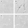
FIG. 1
FIG. 1
Histochemical staining of Arf-expressing p21−/− and p21+/+ cells for SA-β-gal activity. Cells were infected with pBabe-puro-Arf or empty pBabe-puro vectors, selected with puromycin for 5 days, stained for SA-β-gal activity, and photographed. (A) p21+/+ cells, pBabe-puro. (B) p21+/+ cells, pBabe-puro-Arf. (C) p21−/− cells, pBabe-puro. (D) p21−/− cells, pBabe-puro-Arf. All panels are shown at equal magnification.
FIG. 2
FIG. 2
BrdU incorporation assays of Arf-expressing p21−/− and p21+/+ cells. (A) Nonimmortalized p21+/+ and p21−/− cells. (B) hTERT-immortalized p21+/+ and p21−/− cells. Cells were treated as indicated in Fig. 1. At the end of the drug selection, cells were labeled for 24 h with BrdU and subsequently processed for histochemical in situ detection of BrdU incorporation. Since hTERT-immortalized cells are puromycin resistant, the pWZL-Blast vector was used in panel B. Cells were scored microscopically in random fields; a minimum of 200 cells were scored for each determination. The percent means and standard deviations of BrdU-positive cells are presented.
FIG. 3
FIG. 3
Immunoblot analysis of Arf-expressing p21+/+ and p21−/− cells. Exponentially growing cells were infected with pBabe-puro-Arf or empty pBabe-puro viruses, and puromycin-resistant pools of cells were selected as indicated in Fig. 1. For each infection, 12 10-cm-diameter dishes were harvested, pooled, and processed for immunoblotting. (A) Arf. (B) p53. (C) p21. (D) p16. (E) Rb. (F) Actin.
FIG. 4
FIG. 4
Immunoblot analysis of Ras-expressing p21+/+ and p21−/− cells. Exponentially growing cells were infected with pBabe-puro-Ha-Ras(G12) or empty pBabe-puro viruses and processed as indicated in Fig. 3. (A) Ras. (B) p53. (C) p16. (D) p21. (E) Rb. (F) Actin.
FIG. 5
FIG. 5
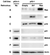
Immunoblot analysis of Arf- or Ras-expressing p53−/− cells. Exponentially growing cells were infected with pBabe-bleo-Arf, pBabe-bleo-Ras, or empty pBabe-bleo viruses, and bleomycin-resistant pools were selected and processed as indicated in Fig. 3. Uninfected p53+/+ cells were included as controls for the immunoblotting detection of p21, Mdm2, and p53. To facilitate this analysis, p53−/− cells immortalized by the expression of hTERT were used. (A) Ras. (B) Arf. (C) p16. (D) p21. (E) Mdm2. (F) p53. (G) Actin.
FIG. 6
FIG. 6
Absence of Arf induction by Ras in normal human fibroblasts. Exponentially growing cells were infected with pBabe-puro-Arf, pBabe-puro-Ras, or empty pBabe-puro viruses and processed as indicated in Fig. 3. (A, B, and C) Immunoblotting analysis of Ras, Arf, and p21, respectively. Two different cell strains of normal human fibroblasts were used: LF1 (left panels) and IMR-90 (right panels). (D and E) RT-PCR analysis of Arf expression in LF1 cells. Quantitative RT-PCR was performed as indicated in Materials and Methods. In the experiment shown, the Arf (D) and β-actin (E) reactions were amplified for 20 and 32 cycles, respectively.
FIG. 7
FIG. 7
Immunoblot analysis of normal human fibroblasts during replicative aging. Human fibroblasts (LF1 cell line) were serially subcultured from early passage until they acquired the senescent phenotype. Protein extracts were prepared at the indicated times: early passage, p14; mid-passage, p28; late passage, p37; senescence, p44. U2OS and HeLa cells were included as negative and positive controls for the immunoblotting detection of Arf. (A) Arf. (B) Mdm2. (C) p53. (D) p16. (E) p21. (F) Rb.
FIG. 8
FIG. 8
Absence of Arf induction during replicative aging of normal human fibroblasts. (A and B) RT-PCR analysis of Arf expression in LF1 cells. In the experiment shown, the Arf (A) and β-actin (B) reactions were amplified for 20 and 32 cycles, respectively. (C) Northern hybridization analysis of Arf and p21 expression in LF1 cells. Data were quantified by PhosphorImager analysis and normalized to a GAPDH signal. Northern hybridization was performed on two separate occasions with samples prepared from cells that were independently passaged into senescence. Both experiments yielded consistent results; one representative experiment is shown. Cells were grown in a 2% O2 atmosphere.
FIG. 9
FIG. 9
Absence of Arf induction during replicative aging of normal human fibroblasts. (A) RNase protection analysis of LF1 cells. (B) RNase protection analysis of WI-38 cells grown under normoxic (lanes 4, 5, and 6) and hypoxic (lanes 7 and 8) conditions. m.w., molecular weight.
FIG. 10
FIG. 10
Summary of p53-p21 and p16-Rb senescence pathways in human and rodent fibroblasts. (A) Replicative senescence. Only human-specific pathways are shown, since the existence of a non-crisis arrest due to telomere attrition has not been convincingly demonstrated in the mouse. (B) Induced (or premature) senescence states. For simplicity, only Ras-mediated oncogenic activation and Arf-mediated “culture shock” (63) are shown, although a number of other effectors and/or stimuli are known to be capable of inducing senescence-like arrest. Common and human- and mouse-specific pathways are shown.

References

    1. Alcorta D A, Xiong Y, Phelps D, Hannon G, Beach D, Barrett J C. Involvement of the cyclin-dependent kinase inhibitor p16(INK4a) in replicative senescence of normal human fibroblasts. Proc Natl Acad Sci USA. 1996;93:13742–13747. - PMC - PubMed
    1. Artandi S E, DePinho R A. Mice without telomerase: what can they teach us about human cancer? Nat Med. 2000;6:852–855. - PubMed
    1. Atadja P, Wong H, Garkavtsev I, Veillette C, Riabowol K. Increased activity of p53 in senescing fibroblasts. Proc Natl Acad Sci USA. 1995;92:8348–8352. - PMC - PubMed
    1. Bates S, Phillips A C, Clark P A, Stott F, Peters G, Ludwig R L, Vousden K H. p14ARF links the tumour suppressors RB and p53. Nature. 1998;395:124–125. - "VSports最新版本" PubMed
    1. Bayreuther K, Rodemann H P, Hommel R, Dittmann K, Albiez M, Francz P I. Human skin fibroblasts in vitro differentiate along a terminal cell lineage. Proc Natl Acad Sci USA. 1988;85:5112–5116. - PMC - PubMed

VSports手机版 - Publication types

MeSH terms