Skip to main page content
U.S. flag

An official website of the United States government

Dot gov

The . gov means it’s official. Federal government websites often end in VSports app下载. gov or . mil. Before sharing sensitive information, make sure you’re on a federal government site. .

Https

The site is secure. The https:// ensures that you are connecting to the official website and that any information you provide is encrypted and transmitted securely. V体育官网.

. 2001 Sep;108(5):679-88.
doi: 10.1172/JCI12976.

Targeted deletion of CX(3)CR1 reveals a role for fractalkine in cardiac allograft rejection

Affiliations

Targeted deletion of CX(3)CR1 reveals a role for fractalkine in cardiac allograft rejection

C A Haskell et al. J Clin Invest. 2001 Sep.

Abstract

Fractalkine (Fk) is a structurally unusual member of the chemokine family. To determine its role in vivo, we generated mice with a targeted disruption of CX(3)CR1, the receptor for Fk. CX(3)CR1(-/-) mice were phenotypically indistinguishable from wild-type mice in a pathogen-free environment. In response to antibody-induced glomerulonephritis, CX(3)CR1(-/-) and CX(3)CR1(+/+) mice had similar levels of proteinuria and injury. CX(3)CR1(-/-) and CX(3)CR1(+/+) mice also developed similar levels of disease in myelin oligodendrocyte glycoprotein-induced experimental autoimmune encephalomyelitis. We performed heterotopic MHC class I/II cardiac transplants from BALB/c mice into C57BL/6 mice VSports手机版. In the absence of cyclosporin A (CsA), there was no difference in graft survival time between CX(3)CR1(-/-) and CX(3)CR1(+/+) recipient mice. However, in the presence of subtherapeutic levels of CsA, graft survival time was significantly increased in the CX(3)CR1(-/-) mice. Characterization of cells infiltrating the grafts revealed a selective reduction in natural killer cells in the CX(3)CR1(-/-) recipients in the absence of CsA and a reduction in macrophages, natural killer cells, and other leukocytes in the presence of CsA. We conclude that Fk plays an important role in graft rejection. The development of CX(3)CR1 antagonists may allow reductions in the doses of immunosuppressive drugs used in transplantation. .


Figures

Figure 1
Figure 1
Targeting vector for the CX3CR1 gene. The top line shows a partial restriction map of the CX3CR1 locus indicating the recognition sites for HindIII (H) and PstI (P). The targeting vector is shown in the center, and the predicted structure of the targeted allele after homologous recombination is shown below. The coding region for CX3CR1 is indicated by the thick bar. The neo gene is flanked by loxP sites for removal. Also indicated are the locations of the PCR primers and the probe used for screening the ES cells and the mice.
Figure 2
Figure 2
Targeted deletion of the CX3CR1 gene. Genomic DNA from F2 offspring of heterozygous (+/–) parents was screened by both Southern blot analysis and PCR. (a) Southern blot. Restriction endonuclease digestion with HindIII was probed with a fragment from the 5′ untranslated region and produced a wild-type fragment of 8 kb and a targeted fragment of 3.1 kb. The wild-type and targeted fragments are indicated with the genotype of each mouse shown above the lane. (b) PCR. A set of three primers was used: one 5′ primer that annealed in the 5′ untranslated region and two 3′ primers, one each from the neo and CX3CR1 genes. The reaction resulted in band sizes of 700 bp for the wild-type allele and 500 bp for the targeted allele.
Figure 3
Figure 3
Peripheral blood cell adhesion to Fk. Peripheral blood was obtained from CX3CR1 wild-type and null mice, and the leukocytes were isolated as a buffy coat. Cells were labeled with the fluorescent dye 2′,7′-bis-(2-carboxyethyl)-5-(and-6)-carboxyfluorescein, acetoxymethyl ester, and tested for adhesion to Fk in cell culture chambers. Excess cells were washed off after a 30-minute incubation, and adhesion was measured as a function of fluorescence. Results are reported as arbitrary fluorescent units. Excess soluble Fk (100 nM) was added to compete with adhesion to the tethered Fk. This assay is representative of three performed. The adhesion of wild-type cells was significantly reduced by the addition of soluble Fk (P < 0.001).
Figure 4
Figure 4
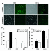
Cell types adhering to Fk. (a) Peripheral blood leukocytes from CX3CR1+/+ mice were isolated as a buffy coat and either spun onto a glass slide (cytospin) or allowed to adhere to Ab- tethered Fk (Fk-adherent). Cells were then labeled with biotinylated Ab’s (CD3 or CD11b) and fluorescent streptavidin for identification. Bright-field and fluorescent images were captured. (b) Quantitation of cell types binding to Fk. Three fields were counted for each labeling condition, and the percentage of each cell type in the input population and Fk-adherent population is shown. The experiment shown is representative of two. (c) Adhesion of purified NK and T cells to Fk. NK (NK1.1+, CD3) and T (CD3+, NK1.1) cells were purified and fluorescently labeled. Cells were allowed to adhere to Ab-tethered Fk, in the presence or absence of soluble (100 nM) Fk. The experiment shown is representative of three. The asterisk indicates P < 0.005.
Figure 5
Figure 5
Histology of control and nephritic kidneys. CX3CR1+/+ (a, c) and CX3CR1–/– (b, d) mice were preimmunized with normal sheep serum and then injected intravenously with normal sheep serum or nephrotoxic serum. Sections of kidneys harvested from mice immunized with normal sheep serum show normal renal morphology (a, b). In contrast, cortical sections obtained 21 days after injection with nephrotoxic serum (c, d) show proliferative and inflammatory glomerular changes, including hypercellularity, focal necrosis, increased matrix, thickening of capillary loops, and occlusion by matrix and microthrombi, as well as a periglomerular interstitial mononuclear cell infiltrate. (H&E-stained paraffin sections; original magnification, ×790). Shown are representative sections from 4 to 6 animals per group.
Figure 6
Figure 6
Lack of requirement for CX3CR1 expression in development of murine EAE. Wild-type and CX3CR1-null mice were immunized with MOG peptide and weighed and scored daily. No significant differences were noted at any point. Results are representative of two separate experiments, using 10 mice per group in each experiment.
Figure 7
Figure 7
Expression of Fk and CX3CR1 during development of cardiac allograft rejection. (a) RPA analysis of Fk mRNA expression showing baseline expression in two control hearts (day 0) and increasing expression in allografts harvested at day 1, 3, or 7 after transplant. Arrow indicates Fk-protected fragment. Each lane represents mRNA from a different allograft. (b) Immunoperoxidase staining of normal heart with an anti-Fk Ab. Heart harvested 7 days after transplant and stained with an anti-Fk Ab (c) or with control IgG (d). (e) Infiltrating mononuclear cells in day-7 allografts stained for CX3CR1. Arrows in be indicate examples of intragraft vascular endothelium; cryostat sections with hematoxylin counterstain. Original magnifications, ×200.
Figure 8
Figure 8
Survival of CX3CR1–/– and CX3CR1+/+ recipient mice after cardiac transplantation. CX3CR1–/– mice rejected fully MHC-disparate cardiac allografts at the same tempo as CX3CR1+/+ recipients. However, whereas administration of a subtherapeutic dose of CsA prolonged allograft survival only 2–3 days in CX3CR1+/+ recipients, allograft survival was significantly prolonged in CX3CR1–/– mice treated with the same course of CsA (P < 0.005 Mann-Whitney vs. wild-type/CsA, n = 6/group).
Figure 9
Figure 9
Comparison of histology of cardiac allografts. (a) CX3CR1+/+ recipients, (c) CX3CR1+/+ recipients treated with CsA, and (b) CX3CR1–/– recipients each show severe cellular rejection with extensive mononuclear cell infiltration and myocyte necrosis (arrows). CX3CR1–/– recipients (b) also showed areas of coagulative necrosis (arrowhead). By contrast, CX3CR1–/– recipients (d) treated with CsA showed intact myocardium and vessels and only a mild degree of mononuclear cell infiltration. (H&E-stained paraffin sections, representative of n = 6/group).
Figure 10
Figure 10
Serial analysis of NK cell and macrophage infiltration of cardiac allografts. Allografts (3/group) were harvested on day 0 (pretransplant) and 1, 3, and 7 days after transplant, and the extent (mean ± SD, 20 fields/graft) of NK cell (DX3+) and macrophage (F4/80+) infiltration was determined. (a) The number of intragraft NK cells peaked at day 3 and was significantly decreased on days 1 and 3 in the CX3CR1–/– recipients. Administration of CsA decreased NK cell infiltration in both CX3CR1+/+ and CX3CR1–/– mice (*P < 0.001, ** P < 0.005). On day 3 after transplant in the presence of CsA there were fewer NK cells in the CX3CR1–/– recipients than in the CX3CR1+/+ mice (P < 0.05). (b) Macrophage recruitment at days 3 and day 7 after transplant was diminished by CsA (*P < 0.001 comparing no CsA versus plus CsA). Macrophage recruitment was most depressed in CX3CR1–/– recipients in the presence of CsA (P < 0.01 comparing CX3CR1+/+ versus CX3CR1–/– recipients at day 3 after transplant; P < 0.001 comparing these same groups at day 7).
Figure 11
Figure 11
Leukocyte accumulation at day 7 after transplant. Analysis of immunoperoxidase-stained cell populations showed significant reductions (*P < 0.01) in recruitment of intragraft CD45+ cells (all leukocytes); T cells (CD3); CD4 and CD8 T cell subsets; macrophages and IL-2R+ (CD25+) cells in CX3CR1–/– animals treated with CsA vs. each of the other three groups. (Mean ± SD, 20 consecutive fields per graft and three grafts per group.)
Figure 12
Figure 12
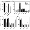
Chemokine and cytokine expression in rejecting allografts. Analysis of day 7 cardiac allografts by RNase protection. (a) Use of CsA did not affect intragraft expression of Fk mRNA, though overall levels in CX3CR1–/– were decreased (P < 0.01) compared with CX3CR1+/+ recipients. (b) Compared with CX3CR1+/+ recipients, targeting of CX3CR1 plus CsA resulted in significant decreases in the chemokines MIP-1α, MIP-1β, RANTES, IP-10, and MCP-1. Ltn, lymphotoxin (c) Decreased levels of IFN-γ and IL-6 in CX3CR1–/– mice treated with CsA. (d) Reduced expression of CCR1 and CCR5 in CX3CR1–/– mice treated with CsA. (Mean ± SD, n = 3/group, Mann-Whitney U test, *P < 0.05, **P < 0.01, and ***P < 0.005.)

References (VSports app下载)

    1. Bazan JF, et al. A new class of membrane-bound chemokine with a CX3C motif. Nature. 1997;385:640–644. - PubMed
    1. Imai T, et al. Identification and molecular characterization of fractalkine receptor CX3CR1, which mediates both leukocyte migration and adhesion. Cell. 1997;91:521–530. - "VSports" PubMed
    1. Haskell CA, Cleary MD, Charo IF. Molecular uncoupling of fractalkine-mediated cell adhesion and signal transduction. Rapid flow arrest of CX3CR1-expressing cells is independent of G-protein activation. J Biol Chem. 1999;274:10053–10058. - PubMed
    1. Fong AM, et al. Fractalkine and CX3CR1 mediate a novel mechanism of leukocyte capture, firm adhesion, and activation under physiologic flow. J Exp Med. 1998;188:1413–1419. - VSports手机版 - PMC - PubMed
    1. Matloubian M, David A, Engel S, Ryan JE, Cyster JG. A transmembrane CXC chemokine is a ligand for HIV-coreceptor Bonzo. Nat Immunol. 2000;1:298–304. - PubMed

Publication types

MeSH terms