Skip to main page content
U.S. flag

An official website of the United States government

Dot gov

The VSports app下载. gov means it’s official. Federal government websites often end in . gov or . mil. Before sharing sensitive information, make sure you’re on a federal government site. .

Https

The site is secure. The https:// ensures that you are connecting to the official website and that any information you provide is encrypted and transmitted securely V体育官网. .

. 2001 Aug 20;194(4):393-406.
doi: 10.1084/jem.194.4.393.

Selective abrogation of major histocompatibility complex class II expression on extrahematopoietic cells in mice lacking promoter IV of the class II transactivator gene

Affiliations

Selective abrogation of major histocompatibility complex class II expression on extrahematopoietic cells in mice lacking promoter IV of the class II transactivator gene

"VSports最新版本" J M Waldburger et al. J Exp Med. .

VSports手机版 - Abstract

MHC class II (MHCII) molecules play a pivotal role in the induction and regulation of immune responses. The transcriptional coactivator class II transactivator (CIITA) controls MHCII expression. The CIITA gene is regulated by three independent promoters (pI, pIII, pIV). We have generated pIV knockout mice. These mice exhibit selective abrogation of interferon (IFN)-gamma-induced MHCII expression on a wide variety of non-bone marrow-derived cells, including endothelia, epithelia, astrocytes, and fibroblasts VSports手机版. Constitutive MHCII expression on cortical thymic epithelial cells, and thus positive selection of CD4(+) T cells, is also abolished. In contrast, constitutive and inducible MHCII expression is unaffected on professional antigen-presenting cells, including B cells, dendritic cells, and IFN-gamma-activated cells of the macrophage lineage. pIV(-/-) mice have thus allowed precise definition of CIITA pIV usage in vivo. Moreover, they represent a unique animal model for studying the significance and contribution of MHCII-mediated antigen presentation by nonprofessional antigen-presenting cells in health and disease. .


Figures

Figure 1
Figure 1
Cell-specific and inducible MHCII expression is controlled by alternative usage of three promoters (pI, pIII, and pIV) of the Mhc2ta gene. pI and pIII are primarily active in DCs and B cells, respectively, while pIV is activated by IFN-γ. Usage of these promoters leads to the transcription (arrows) and splicing of alternative first exons (open boxes) to a shared second exon (shaded boxes). Incorporation of exons I and III leads to the synthesis of type I and type III CIITA mRNA encoding CIITA proteins having specific NH2-terminal extensions of 94 and 17 amino acids, respectively. In contrast, translation of type IV CIITA is initiated at the AUG found in the shared second exon. The three different forms of CIITA mRNA can be distinguished in RNase protection assay (rpa) with the indicated probes. CIITA activates transcription of MHCII genes by associating with proteins (RFX, X2BP, and NF-Y) that bind to MHCII promoters. ut, 5′ untranslated region.
Figure 2
Figure 2
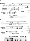
Generation of Mhc2ta pIV−/− mice. (A) The regulatory region of the wild type Mhc2ta locus, the targeting construct, and the targeted locus are depicted. Exons I, III, and IV are represented as open boxes, loxP sites (P) are shown as filled triangles, and the neo gene is indicated by an open arrow. HindIII sites (H) used for Southern blotting are indicated. A representative blot for a positive ES cell clone is shown below: the 3′ external probe (shown below the wild-type locus) hybridizes to a 10-kb fragment in the wild-type locus and to a 9-kb fragment in the targeted locus. (B) Cre-mediated deletion of the targeted locus. The wild-type, targeted, and deleted loci are depicted as in A. Arrows labeled f1, r1, f 2, and r2 represent PCR primers used for genotyping of mice (see C). BamHI sites (B) used for Southern blotting are indicated. A representative blot for heterozygous mice is shown below: the internal probe (shown below the wild-type locus) hybridizes to a 3.4-kb fragment in the wild-type locus, to a 2.6-kb fragment in the targeted locus, and to a 1-kb fragment in the deleted locus. λDNA digested with StyI was used as a molecular mass marker. (C) Genotyping of mice was done by PCR using the primer pair f1+r1 to amplify a 500-bp fragment specific for the wild-type locus and the primer pair f 2+r2 to amplify a 700-bp fragment diagnostic for the deleted locus. A representative experiment is shown for the offspring derived from a cross between two heterozygous pIV+/− mice. (D) Type IV CIITA mRNA is not induced in pIV−/− mice. MEF isolated from heterozygous (+/−) and homozygous (−/−) mice were cultivated in the absence (lanes 1 and 3) or presence (lanes 2 and 4) of IFN-γ. The presence of type IV CIITA mRNA was assayed by RNase protection using the type IV specific probe (Fig. 1). A GAPDH probe was used as internal control. (E) Expression of types I and III CIITA mRNA is not affected by the deletion of pIV. Bone marrow–derived DCs generated from control littermates (+/−) and homozygous mutants (−/−) were analyzed by RNase protection using the type I–specific probe (top). Total spleen RNA from pIV+/− and pIV−/− animals was analyzed using the type III–specific probe (bottom). A TATA box binding protein (TBP) probe was used as internal control.
Figure 2
Figure 2
Generation of Mhc2ta pIV−/− mice. (A) The regulatory region of the wild type Mhc2ta locus, the targeting construct, and the targeted locus are depicted. Exons I, III, and IV are represented as open boxes, loxP sites (P) are shown as filled triangles, and the neo gene is indicated by an open arrow. HindIII sites (H) used for Southern blotting are indicated. A representative blot for a positive ES cell clone is shown below: the 3′ external probe (shown below the wild-type locus) hybridizes to a 10-kb fragment in the wild-type locus and to a 9-kb fragment in the targeted locus. (B) Cre-mediated deletion of the targeted locus. The wild-type, targeted, and deleted loci are depicted as in A. Arrows labeled f1, r1, f 2, and r2 represent PCR primers used for genotyping of mice (see C). BamHI sites (B) used for Southern blotting are indicated. A representative blot for heterozygous mice is shown below: the internal probe (shown below the wild-type locus) hybridizes to a 3.4-kb fragment in the wild-type locus, to a 2.6-kb fragment in the targeted locus, and to a 1-kb fragment in the deleted locus. λDNA digested with StyI was used as a molecular mass marker. (C) Genotyping of mice was done by PCR using the primer pair f1+r1 to amplify a 500-bp fragment specific for the wild-type locus and the primer pair f 2+r2 to amplify a 700-bp fragment diagnostic for the deleted locus. A representative experiment is shown for the offspring derived from a cross between two heterozygous pIV+/− mice. (D) Type IV CIITA mRNA is not induced in pIV−/− mice. MEF isolated from heterozygous (+/−) and homozygous (−/−) mice were cultivated in the absence (lanes 1 and 3) or presence (lanes 2 and 4) of IFN-γ. The presence of type IV CIITA mRNA was assayed by RNase protection using the type IV specific probe (Fig. 1). A GAPDH probe was used as internal control. (E) Expression of types I and III CIITA mRNA is not affected by the deletion of pIV. Bone marrow–derived DCs generated from control littermates (+/−) and homozygous mutants (−/−) were analyzed by RNase protection using the type I–specific probe (top). Total spleen RNA from pIV+/− and pIV−/− animals was analyzed using the type III–specific probe (bottom). A TATA box binding protein (TBP) probe was used as internal control.
Figure 3
Figure 3
Selective loss of IFN-γ–induced MHCII expression on non-bone marrow–derived cells in pIV-deficient mice. (A) MEFs from pIV−/− fetuses exhibit a selective loss of IFN-γ–induced MHCII expression. MEFs isolated from wild-type (+/+), heterozygous (+/−), and homozygous (−/−) mice were treated with IFN-γ and analyzed by two-color FACS® for the induction of MHCI and MHCII expression. (B) The deletion of pIV does not eliminate IFN-γ–induced MHCII expression in macrophages. Thioglycollate-elicited peritoneal macrophages were isolated from heterozygous (+/−) and homozygous (−/−) mice, cultured in the absence (open profile) or presence (filled profile) of IFN-γ, and then analyzed by FACS® for MHCII expression. (C) IFN-γ–induced MHCII expression is lost in astrocytes but retained in microglia from pIV-deficient mice. Brain-derived cells from pIV−/− mice were cultured in the absence (open profiles) or presence (solid profiles) of IFN-γ. CD11b+ microglial cells (R2, top right) and CD11b astrocytes (R1, center right) were analyzed by FACS® for expression of MHCII. Activation of the astrocytes by IFN-γ was verified by examining the upregulation of MHCI expression (bottom right).
Figure 4
Figure 4
IFN-γ–induced activation of Mhc2ta expression is mediated largely by pI. Thioglycollate-elicited peritoneal macrophages from control (+/−) or homozygous mutant (−/−) mice were cultured in the absence (lanes 1 and 3) or presence (lanes 2 and 4) of IFN-γ. Expression of CIITA was analyzed by RNase protection using probes specific for type IV (A), type I (B), and type III (C) mRNA. The band corresponding to the specific mRNA (I, III, or IV) and the band representing mRNA derived from the other two promoters (NonI, NonIII, and NonIV) are indicated. A probe for GAPDH mRNA was used as internal control. The percentages of total CIITA mRNA corresponding to types I, III, and IV were quantified by PhosphorImager analysis of RNase protection experiments similar to those shown in A–C (D).
Figure 4
Figure 4
IFN-γ–induced activation of Mhc2ta expression is mediated largely by pI. Thioglycollate-elicited peritoneal macrophages from control (+/−) or homozygous mutant (−/−) mice were cultured in the absence (lanes 1 and 3) or presence (lanes 2 and 4) of IFN-γ. Expression of CIITA was analyzed by RNase protection using probes specific for type IV (A), type I (B), and type III (C) mRNA. The band corresponding to the specific mRNA (I, III, or IV) and the band representing mRNA derived from the other two promoters (NonI, NonIII, and NonIV) are indicated. A probe for GAPDH mRNA was used as internal control. The percentages of total CIITA mRNA corresponding to types I, III, and IV were quantified by PhosphorImager analysis of RNase protection experiments similar to those shown in A–C (D).
Figure 5
Figure 5
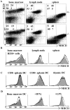
Deletion of Mhc2ta pIV does not affect constitutive MHCII expression in bone marrow–derived cells and secondary lymphoid tissues. (A) Cells isolated from the bone marrow, lymph nodes, and spleens of pIV−/− mice and pIV+/− littermate controls were analyzed by two-color FACS® for the expression of MHCII and the B cell marker B220. The percentage of cells in the four quadrants is indicated for each panel. (B) FACS® analysis of MHCII expression on ex vivo B cells isolated from the bone marrow, lymph nodes, and spleens of pIV−/− mice (solid profiles) and control littermates (open profiles). (C) MHCII expression on ex vivo DCs isolated from pIV−/− mice (solid profiles) and control pIV+/− littermates (open profiles) was examined by four-color FACS® analysis. Cells studied were thymic (CD11c+B220) DCs and splenic DCs of the myeloid (CD11c+B220CD8) and lymphoid (CD11c+B220CD8+) subsets. (D) MHCII expression was analyzed by FACS® on CD11c+ bone marrow–derived DCs from pIV−/− mice (solid profiles) and control littermates (open profiles). The DCs were analyzed as such, after induction with IFN-γ, or after treatment with LPS to induce maturation. (E) Sections of the spleen and Peyer's patches from adult pIV−/− mice and control pIV+/− littermates were stained for MHCII expression (brown color).
Figure 5
Figure 5
Deletion of Mhc2ta pIV does not affect constitutive MHCII expression in bone marrow–derived cells and secondary lymphoid tissues. (A) Cells isolated from the bone marrow, lymph nodes, and spleens of pIV−/− mice and pIV+/− littermate controls were analyzed by two-color FACS® for the expression of MHCII and the B cell marker B220. The percentage of cells in the four quadrants is indicated for each panel. (B) FACS® analysis of MHCII expression on ex vivo B cells isolated from the bone marrow, lymph nodes, and spleens of pIV−/− mice (solid profiles) and control littermates (open profiles). (C) MHCII expression on ex vivo DCs isolated from pIV−/− mice (solid profiles) and control pIV+/− littermates (open profiles) was examined by four-color FACS® analysis. Cells studied were thymic (CD11c+B220) DCs and splenic DCs of the myeloid (CD11c+B220CD8) and lymphoid (CD11c+B220CD8+) subsets. (D) MHCII expression was analyzed by FACS® on CD11c+ bone marrow–derived DCs from pIV−/− mice (solid profiles) and control littermates (open profiles). The DCs were analyzed as such, after induction with IFN-γ, or after treatment with LPS to induce maturation. (E) Sections of the spleen and Peyer's patches from adult pIV−/− mice and control pIV+/− littermates were stained for MHCII expression (brown color).
Figure 6
Figure 6
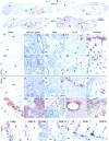
Deletion of Mhc2ta pIV results in a selective loss of IFN-γ–induced MHCII expression on non-bone marrow–derived cells. Sections of newborn mice were stained for MHCII expression (brown color). (Top) Whole body sections of newborn pIV−/− mice (A and C) and control pIV+/− littermates (B and D) that had been injected (C and D) or had not been injected (A and B) with IFN-γ. Positions of the brain (b), lungs (lu), liver (li), intestines (i), and salivary glands (sg) are indicated. (1A–5D) Higher magnifications of representative regions of organs from the mice shown in A–D. Brain sections (1A–1D): m, meninges; p, parenchyma. Submandibular salivary gland sections (2A–2D): d, secretory duct. Lung sections (3A–3D): v, pulmonary veins; b, bronchioles; rb, respiratory bronchiole; bs, bronchus. Liver sections (4A–4D): v, central vein; d, bile duct. Sections through the intestines (5A–5D) show the visceral peritoneum, crypts, and villi. (Bottom) Adjacent sections from the brain (1C), lung (3C), and intestine (5C) of IFN-γ–injected pIV−/− mice were stained for expression of MHCII and a macrophage-specific marker (F4/80).
Figure 7
Figure 7
Deletion of Mhc2ta pIV results in the loss of MHCII expression on cTECs and leads to impaired positive selection of CD4+ T cells. (A) Thymic sections from pIV−/− mice and pIV+/− littermate controls were stained (brown color) for expression of MHCII, the DC marker CD11c, or the macrophage marker F4/80. Regions corresponding to the cortex (c) and medulla (m) are indicated. (B) T cell populations in the thymus, lymph nodes, and spleen of pIV−/− mice and pIV+/− littermate controls were analyzed by FACS® for expression of CD8 and CD4. The percentages of single-positive CD4, single-positive CD8, and double-positive cells are indicated. (C) The ability to generate a primary T cell–dependent antibody response was examined in pIV−/− mice, in pIV+/− littermates, and in negative controls (CIITA−/− and I-Aα2/− mice). High-affinity IgG titers produced after immunization with NP-CGG were measured at the indicated dilutions of the sera.
Figure 7
Figure 7
Deletion of Mhc2ta pIV results in the loss of MHCII expression on cTECs and leads to impaired positive selection of CD4+ T cells. (A) Thymic sections from pIV−/− mice and pIV+/− littermate controls were stained (brown color) for expression of MHCII, the DC marker CD11c, or the macrophage marker F4/80. Regions corresponding to the cortex (c) and medulla (m) are indicated. (B) T cell populations in the thymus, lymph nodes, and spleen of pIV−/− mice and pIV+/− littermate controls were analyzed by FACS® for expression of CD8 and CD4. The percentages of single-positive CD4, single-positive CD8, and double-positive cells are indicated. (C) The ability to generate a primary T cell–dependent antibody response was examined in pIV−/− mice, in pIV+/− littermates, and in negative controls (CIITA−/− and I-Aα2/− mice). High-affinity IgG titers produced after immunization with NP-CGG were measured at the indicated dilutions of the sera.

Comment in

References

    1. Cresswell P. Assembly, transport, and function of MHC class II molecules. Annu. Rev. Immunol. 1994;12:259–293. - PubMed
    1. Viret C., Janeway C.A. MHC and T cell development. Rev. Immunogenet. 1999;1:91–104. - PubMed
    1. Harton J.A., Ting J.P. Class II transactivatormastering the art of major histocompatibility complex expression. Mol. Cell Biol. 2000;20:6185–6194. - PMC (V体育2025版) - PubMed
    1. Boss J.M. Regulation of transcription of MHC class II genes. Curr. Opin. Immunol. 1997;9:107–113. - PubMed
    1. Reith W., Mach B. The bare lymphocyte syndrome and the regulation of MHC expression. Annu. Rev. Immunol. 2001;19:331–373. - PubMed

"VSports" MeSH terms