Skip to main page content
U.S. flag

An official website of the United States government

Dot gov

The . gov means it’s official. Federal government websites often end in . gov or . mil. Before sharing sensitive information, make sure you’re on a federal government site VSports app下载. .

Https

The site is secure V体育官网. The https:// ensures that you are connecting to the official website and that any information you provide is encrypted and transmitted securely. .

. 2001 Aug;75(16):7252-65.
doi: 10.1128/JVI.75.16.7252-7265.2001.

V体育官网入口 - Human immunodeficiency virus type 1 Vif protein is packaged into the nucleoprotein complex through an interaction with viral genomic RNA

Affiliations

V体育平台登录 - Human immunodeficiency virus type 1 Vif protein is packaged into the nucleoprotein complex through an interaction with viral genomic RNA

M A Khan et al. J Virol. 2001 Aug.

Abstract

The human immunodeficiency virus type 1 (HIV-1) Vif protein plays a critical role in the production of infectious virions. Previous studies have demonstrated the presence of small amounts of Vif in virus particles. However, Vif packaging was assumed to be nonspecific, and its functional significance has been questioned. We now report that packaging of Vif is dependent on the packaging of viral genomic RNA in both permissive and restrictive HIV-1 target cells. Mutations in the nucleocapsid zinc finger domains that abrogate packaging of viral genomic RNA abolished packaging of Vif. Additionally, an RNA packaging-defective virus exhibited significantly reduced packaging of Vif. Finally, deletion of a putative RNA-interacting domain in Vif abolished packaging of Vif into virions. Virion-associated Vif was resistant to detergent extraction and copurified with components of the viral nucleoprotein complex and functional reverse transcription complexes. Thus, Vif is specifically packaged into virions as a component of the viral nucleoprotein complex VSports手机版. Our data suggest that the specific association of Vif with the viral nucleoprotein complex might be functionally significant and could be a critical requirement for infectivity of viruses produced from restrictive host cells. .


Figures

FIG. 1
FIG. 1
Vif does not affect the protein composition of HIV virions. Virus preparations from HeLa cells were produced by transient transfection with pNL4-3 or pNL4-3Vif(−). Viruses from T-cell lines and PBMC were obtained from single-cycle-infected cultures. For single-round infection of T-cell lines and PBMC, virus stocks containing the VSV G protein were produced in HeLa cells by cotransfection of an env-deficient pNL4-3 variant (pNLenv-1) and pHCMV-G as described in Materials and Methods. Cells were infected with concentrated virus stocks for 5 h before residual input virus was removed. As a further precaution against contamination by input virus, virus produced within the first 24 h after infection was discarded. Only virus produced thereafter was used for immunoblot analysis. Virus was normalized for equal reverse transcriptase activity and analyzed by immunoblotting using an HIV-positive patient serum (APS) or an antiserum to integrase (α-Int) (29). Viral proteins are identified on the right.
FIG. 2
FIG. 2
Vif has no discernible effect on synthesis or maturation of viral proteins or on virus release in HIV-infected H9 cells. H9 cells were single-cycle infected with wild-type (NLenv-1) or Vif-defective [NLenv-1/Vif(−)] variants of the env-defective NL4-3 isolate pseudotyped with the VSV-G envelope as described for Fig. 1. Twenty-four hours after infection, cells were metabolically labeled for 1 h with [35S]methionine and chased for up to 4 h. Equal aliquots of cells and supernatants were harvested and subjected to immunoprecipitation with an HIV-positive patient serum. Immunoprecipitated proteins were separated by SDS-PAGE and visualized by fluorography. Proteins are identified on the left.
FIG. 3
FIG. 3
Vif is present in virus preparations derived from nonpermissive H9 cells. H9 cells were infected with NL4-3. Virus was harvested near peak infection, filtered through 0.45-μm filters to remove cellular debris, and concentrated by ultracentrifugation. Concentrated virus stocks were subjected to linear 10 to 50% sucrose gradient centrifugation as described in Materials and Methods. Individual gradient fractions were analyzed by immunoblotting using an HIV-positive patient serum (APS) or antibodies to integrase (α-Int) or Vif (α-Vif). Viral proteins are identified on the right.
FIG. 4
FIG. 4
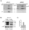
Mutation of the nucleocapsid zinc finger domain abolishes Vif incorporation into virions. (A) HeLa cells were transiently transfected with plasmid DNAs encoding wild-type HIV-1 (pNL4-3) or a nucleocapsid zinc finger mutant of NL4-3 (pDB653). Virus-containing supernatants were harvested 48 h after transfection, concentrated, and subjected to 10 to 60% linear sucrose gradient centrifugation. Individual gradient fractions were collected and subjected to immunoblotting using an HIV-positive patient serum (APS) or a Vif-specific antiserum (α-Vif). (B) HeLa cells were transfected with the Vif expression vector pNL-A1 (lanes a) or contransfected with pNL-A1 plus pNL4-3 (lanes b) or the zinc finger mutant pDB653 (lanes c). Virus-containing supernatants were harvested 48 h after transfection and pelleted through a cushion of 20% sucrose. Cell lysates and viral pelleted fractions were subjected to immunoblot analysis using an HIV-positive patient serum (APS) or a Vif-specific antiserum (α-Vif). (C) Bands corresponding to Vif in panel B were quantified by densitometric scanning, and the proportion of Vif identified in the pooled gradient fractions was calculated as percentage of total intra- and extracellular Vif.
FIG. 5
FIG. 5
Efficient packaging of Vif requires viral RNA. (A) Schematic outline of the structure of the RNA-packaging mutant pC-Help (42). The plasmid lacks both viral LTRs and carries a deletion upstream of gag (indicated by the triangle) which eliminates a putative RNA-packaging signal. In addition, the plasmid carries a deletion in the env gene (indicated by a broken line). polyA, poly(A) addition site. (B) HeLa cells were transfected with pC-Help plasmid DNA. Virus-containing supernatants were harvested 48 h after transfection and concentrated by ultracentrifugation. Concentrated virus was either analyzed directly (lane b) or subjected to sucrose step gradient centrifugation as described in Materials and Methods (lanes c to e). Three equal fractions were collected as indicated in the diagram on the right. Cell lysates (lane a) and viral fractions were separated by SDS–12.5% PAGE and subjected to immunoblot analysis using an HIV-positive patient serum (APS) or a Vif-specific antiserum (α-Vif). Vif-specific bands in lanes a and b were quantified by densitometric scanning as in Fig. 4. Viral proteins are identified on the right.
FIG. 6
FIG. 6
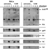
Vif is resistant to detergent extraction of virus derived from HeLa and H9 cells. HeLa-derived virus stocks were prepared by transient transfection of HeLa cells with pNL4-3 DNA as described for Fig. 4. For the preparation of virus stocks from H9 cells, H9 cells were infected with the NL4-3 isolate. Virus-containing supernatants were harvested near the peak of the infection. HeLa- and H9-derived viruses were concentrated by ultracentrifugation and subjected to step gradient purification as described for Fig. 5. To assess the detergent sensitivity of viral components, 50% of the virus preparation was subjected to step gradient centrifugation in the presence of Triton X-100 (X100). The remaining virus was left untreated (untreated). Three fractions containing soluble proteins (fraction 1), a buffer fraction (fraction 2), and the virus-containing fraction (fraction 3) were harvested as in Fig. 5. Individual gradient fractions were subjected to immunoblot analysis using an HIV-positive patient serum (top panels) or antibodies to gp41, integrase (Int), nucleocapsid (NC), or Vif, as indicated on the right.
FIG. 7
FIG. 7
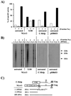
Viral genomic RNA and the reverse transcription complex are insensitive to detergent treatment. (A) Step gradient fractions of virus preparations derived from H9 cells (Fig. 6) or from HeLa cells transfected with pC-Help (Fig. 5B) or pDB653 were examined for reverse transcriptase activity using a conventional RT assay (67). Values are expressed as a percentage of the total reverse transcriptase activity. (B) All fractions analyzed in panel A were subsequently analyzed by an endogenous reverse transcriptase assay as described in Materials and Methods. The predicted size of the tRNALys-derived (−)ssDNA is 253 nucleotides (b). Removal of the tRNA component from the (−)ssDNA by RNase treatment is expected to reduce its size to 181 nucleotides. A synthetic oligonucleotide, Z85, was included in the reactions to control for the presence of unspliced viral genomic RNA in individual fractions. The predicted size of the Z85-primed cDNA product is 354 nucleotides. (C) Schematic outline of the predicted products from the endogenous RT assay. PBS, binding site for the tRNALys primer; SD, splice donor site. A deletion in C-Help eliminating a putative RNA-packaging signal is indicated by a broken line.
FIG. 8
FIG. 8
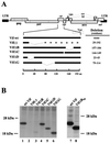
Deletions in Vif do not affect Vif expression levels. (A) Schematic representation of deletions introduced into the vif gene. Construction of the individual mutants is described in Materials and Methods. Deleted regions (amino acid positions) in Vif are denoted on the right. (B) HeLa cells were transfected with pNL-A1 (wt Vif), pNL-A1Vif(−), or individual deletion mutants as indicated. Twenty-four hours after transfection, cells were metabolically labeled for 90 min with [35S]methionine. Cell lysates were subjected to immunoprecipitation with a Vif-specific antibody followed by SDS–12.5% PAGE (lanes 1 to 6). In a similar experiment, cells were transfected with either pNL-A1 (wt Vif) or a Flag epitope-tagged variant of VifΔB, pNL-A1/VifΔBFlag. Cells were labeled as before and precipitated with either a Vif-specific antibody (wt Vif, lane 7) or the epitope tag-specific M2 monoclonal antibody (VifΔBFlag, lane 8). Proteins were subjected to SDS–12.5% PAGE and visualized by fluorography.
FIG. 9
FIG. 9
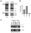
Deletion of a central domain in Vif abolishes packaging into virions. (A) To assess the impact of deletions in Vif on packaging into virions, HeLa cells were cotransfected with pNL4-3Vif(−) and either pNL-A1 (wt Vif), pNL-A1/VifΔD (VifΔD), or pNL-A1/VifΔG (VifΔG). Cells and virus-containing supernatants were harvested 48 h posttransfection. Virions were purified and concentrated by sucrose step gradient centrifugation as described in Materials and Methods. Defined fractions of cell lysates and viral pellets were subjected to SDS–12.5% PAGE followed by immunoblotting with an HIV-positive patient serum (APS) or a Vif-specific antibody (α-Vif). (B) Intracellular and virus-associated Vif proteins detected in panel A were quantified using the FujiX Image Gauge software. The amount of Vif associated with virions was calculated as a percentage of total intra- and extracellular Vif. (C) HeLa cells were transfected with pNL-A1 (wt Vif) or pNL-A1/VifΔG (VifΔG). In addition to the expression of Vif, both plasmids encode authentic viral mRNAs for the expression of Vpr, Tat, Rev, Vpu, Env, and Nef (62). Cells were metabolically labeled for 90 min as described for Fig. 8B. Cells were fractionated into soluble (lanes a), detergent-soluble (lanes b), and detergent-resistant (lanes c) fractions in either the presence or absence of RNase A as described in Materials and Methods. Vif proteins present in individual fractions were precipitated using the Vif-specific polyclonal antibody, separated by SDS–12.5% PAGE, and visualized by fluorography.

References

    1. Adachi A, Gendelman H E, Koenig S, Folks T, Willey R, Rabson A, Martin M A. Production of acquired immunodeficiency syndrome-associated retrovirus in human and nonhuman cells transfected with an infectious molecular clone. J Virol. 1986;59:284–291. - PMC - PubMed
    1. Akari H, Sakuragi J, Takebe Y, Tomonaga K, Kawamura M, Fukasawa M, Miura T, Shinjo T, Hayami M. Biological characterization of human immunodeficiency virus type 1 and type 2 mutants in human peripheral blood mononuclear cells. Arch Virol. 1992;123:157–167. - PubMed
    1. Akari H, Uchiyama T, Fukumori T, Iida S, Koyama A H, Adachi A. Pseudotyping human immunodeficiency virus type 1 by vesicular stomatitis virus G protein does not reduce the cell-dependent requirement of vif for optimal infectivity: functional difference between Vif and Nef. J Gen Virol. 1999;80:2945–2949. - PubMed
    1. Aldovini A, Young R A. Mutations of RNA and protein sequences involved in human immunodeficiency virus type 1 packaging result in production of noninfectious virus. J Virol. 1990;64:1920–1926. - PMC - PubMed
    1. Bess J W, Jr, Gorelick R J, Bosche W J, Henderson L E, Arthur L O. Microvesicles are a source of contaminating cellular proteins found in purified HIV-1 preparations. Virology. 1997;230:134–144. - PubMed (V体育安卓版)

VSports最新版本 - Publication types

MeSH terms (VSports)

LinkOut - more resources (V体育平台登录)