Skip to main page content
U.S. flag

An official website of the United States government

Dot gov

The VSports app下载. gov means it’s official. Federal government websites often end in . gov or . mil. Before sharing sensitive information, make sure you’re on a federal government site. .

Https

The site is secure. The https:// ensures that you are connecting to the official website and that any information you provide is encrypted and transmitted securely V体育官网. .

. 2001 Jul;69(7):4358-65.
doi: 10.1128/IAI.69.7.4358-4365.2001.

CdtA, CdtB, and CdtC form a tripartite complex that is required for cytolethal distending toxin activity

Affiliations

CdtA, CdtB, and CdtC form a tripartite complex that is required for cytolethal distending toxin activity

VSports最新版本 - M Lara-Tejero et al. Infect Immun. 2001 Jul.

"VSports" Abstract

Campylobacter jejuni encodes a cytolethal distending toxin (CDT) that causes cells to arrest in the G(2)/M transition phase of the cell cycle. Highly related toxins are also produced by other important bacterial pathogens VSports手机版. CDT activity requires the function of three genes: cdtA, cdtB, and cdtC. Recent studies have established that CdtB is the active subunit of CDT, exerting its effect as a nuclease that damages the DNA and triggers cell cycle arrest. Microinjection of CdtB into target cells led to G(2)/M arrest and cytoplasmic distention, in a manner indistinguishable from that caused by CDT treatment. Despite this progress, nothing is known about the composition of the CDT holotoxin or the function of CdtA and CdtC. We show here that, when applied individually, purified CdtA, CdtB, or CdtC does not exhibit toxic activity. In contrast, CdtA, CdtB, and CdtC when combined, interact with one another to form an active tripartite holotoxin that exhibits full cellular toxicity. CdtA has a domain that shares similarity with the B chain of ricin-related toxins. We therefore proposed that CDT is a tripartite toxin composed of CdtB as the enzymatically active subunit and of CdtA and CdtC as the heterodimeric B subunit required for the delivery of CdtB. .


Figures

FIG. 1
FIG. 1
Diagram of plasmid constructs expressing M45 epitope-tagged versions of the different Cdt proteins.
FIG. 2
FIG. 2
Purified Cdt proteins. Recombinant Cdt proteins were purified as indicated in Materials and Methods, run on a polyacrylamide gel, and stained with Coomassie blue.
FIG. 3
FIG. 3
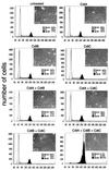
Effect of purified Cdt proteins on cultured intestinal epithelial cells. Purified Cdt proteins alone or in combination (as indicated) were added at a concentration of 10 μg/ml to culture intestinal Henle-407 cells. At 72 h after addition of the proteins, the cells were examined under a phase microscope or processed to measure DNA content by flow cytometry as indicated in Materials and Methods. The scale bar in the microphotographs of cultured intestinal cells treated with the different samples represents 50 μm.
FIG. 4
FIG. 4
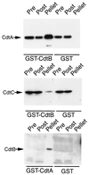
GST pulldown assays for Cdt protein interactions. Whole-cell lysates of E. coli expressing cdt operons encoding M45 epitope-tagged CdtA, CdtB, or CdtC proteins (as indicated to the left of each panel) were probed with purified GST-CdtB, GST-CdtA, or GST (as a negative control) as indicated below each panel. Cdt proteins in the samples before (Pre) and after (Post) the pulldowns or bound to the glutathione-agarose beads (pellet) were detected by Western blotting with a monoclonal antibody directed to the M45 epitope tag. The levels of CdtB in E. coli lysates were not sufficient for its detection in the pre- or postpulldown samples without prior concentration. However, precipitation with GST-CdtA resulted in the concentration of CdtB, which allowed its detection in the sample.
FIG. 5
FIG. 5
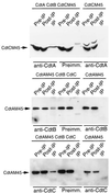
Coimmunoprecipitation assays for Cdt protein interactions. Whole-cell lysates of E. coli expressing different M45 epitope-tagged Cdt proteins in combination with other untagged Cdt proteins (as indicated at the top of each panel) were subjected to coimmunoprecipitation assays with rabbit polyclonal antibodies specific to different Cdt proteins (as indicated below each panel). M45 epitope-tagged Cdt proteins in samples before (Pre-IP) and after (Post-IP) the immunoprecipitation or bound to the protein A-Sepharose beads (pellet) were detected by Western blotting with a monoclonal antibody directed to the M45 epitope. As controls for the specificity of the antibodies, lysates expressing different epitope-tagged Cdt proteins alone (as indicated on the top of the last three lanes of each panel) were treated with the anti-Cdt polyclonal antibodies (as indicated below the last three lanes of each panel), and the immunoprecipitates were analyzed by Western blotting with the M45 monoclonal antibody. Preimm., preimmune serum.
FIG. 6
FIG. 6
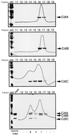
Gel filtration chromatography of Cdt proteins. Purified Cdt proteins, individually or in combination, were loaded onto a Superdex 200 gel filtration column as indicated in Materials and Methods. Then, 1-ml fractions were collected and examined by SDS-PAGE and Coomassie blue staining. Superimposed over the protein bands are the chromatography absorption profiles. Where indicated, the toxicity was examined as described in Materials and Methods using 50 μl of each fraction. The toxicity index represents the inverse of the dilution of each sample that caused visible morphological changes in culture intestinal Henle-407 cells.

References

    1. Collazo C M, Galan J E. Requirement for exported proteins in secretion through the invasion-associated type III system of Salmonella typhimurium. Infect Immun. 1996;64:3524–3531. - PMC (VSports手机版) - PubMed
    1. Comayras C, Tasca C, Peres S Y, Ducommun B, Oswald E, De Rycke J. Escherichia coli cytolethal distending toxin blocks the HeLa cell cycle at the G2/M transition by preventing cdc2 protein kinase dephosphorylation and activation. Infect Immun. 1997;65:5088–5095. - PMC - PubMed
    1. Cope L D, Lumbley S, Latimer J L, Klesney-Tait J, Stevens M K, Johnson L S, Purven M, Munson R S, Jr, Lagergard T, Radolf J D, Hansen E J. A diffusible cytotoxin of Haemophilus ducreyi. Proc Natl Acad Sci USA. 1997;94:4056–4061. - VSports在线直播 - PMC - PubMed
    1. Cortes-Bratti X, Chaves-Olarte E, Lagergard T, Thelestam M. Cellular Internalization of cytolethal distending toxin from Haemophilus ducreyi. Infect Immun. 2000;68:6903–6911. - PMC - PubMed
    1. Cortes-Bratti X, Chaves-Olarte E, Lagergard T, Thelestam M. The cytolethal distending toxin from the chancroid bacterium Haemophilus ducreyi induces cell-cycle arrest in the G2 phase. J Clin Investig. 1999;103:107–115. - PMC - PubMed

MeSH terms

LinkOut - more resources