Skip to main page content
U.S. flag

An official website of the United States government

Dot gov

The . gov means it’s official. Federal government websites often end in . gov or . mil. Before sharing sensitive information, make sure you’re on a federal government site VSports app下载. .

Https

The site is secure V体育官网. The https:// ensures that you are connecting to the official website and that any information you provide is encrypted and transmitted securely. .

Comparative Study
. 2001 Jan 15;20(1-2):165-77.
doi: 10.1093/emboj/20.1.165.

"VSports手机版" Solution structure of the PHD domain from the KAP-1 corepressor: structural determinants for PHD, RING and LIM zinc-binding domains

Affiliations
Comparative Study

"V体育2025版" Solution structure of the PHD domain from the KAP-1 corepressor: structural determinants for PHD, RING and LIM zinc-binding domains

"V体育ios版" A D Capili et al. EMBO J. .

Abstract

Plant homeodomain (PHD) domains are found in >400 eukaryotic proteins, many of which are transcriptional regulators VSports手机版. Naturally occurring point mutations or deletions of this domain contribute to a variety of human diseases, including ATRX syndrome, myeloid leukemias and autoimmune dysfunction. Here we report the first structural characterization of a PHD domain. Our studies reveal that the PHD domain from KAP-1 corepressor binds zinc in a cross-brace topology between anti-parallel ss-strands reminiscent of RING (really interesting new gene) domains. Using a mutational analysis, we define the structural features required for transcriptional repression by KAP-1 and explain naturally occurring, disease-causing mutations in PHD domains of other proteins. From a comparison of this PHD structure with previously reported RING and LIM (Lin11/Isl-1/Mec-3) structures, we infer sequence determinants that allow discrimination among PHD, RING and LIM motifs. .


Figures
None
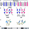
Fig. 1. (A) The consensus sequences that define the PHD, RING and LIM domains. C, cysteine; H, histidine; X, any residue. Above the consensus sequence the number of each metal ligand, ml, is given. The first and second pairs of sequential metal ligands are in dark blue, while the third and fourth pairs are in magenta. (B) Demonstration of zinc ligation patterns found in RING and LIM domains. The RING uses a cross-brace ligation scheme while the LIM uses a sequential ligation scheme. Numbers correlate to the number of the metal ligand as defined in (A). Zinc atoms are represented by gray ovals and the zinc-binding sites are denoted by roman numerals. (C) Ribbon diagrams of the three-dimensional structures. Zinc atoms are represented by spheres. Structures are from the RING of PML (1BOR) and for LIM (1A7I). (D) A schematic representation of the domains in the KAP-1 protein. RING, the RING domain; B1 and B2, two adjacent B-boxes; Coiled-Coil, a leucine coiled-coil domain; hatched box, the TIFF signature sequence; black box, the HP1-binding site; PHD, the PHD domain; Bromo, the bromodomain.
None
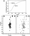
Fig. 2. (A) CD spectrum of untagged KAP-1 PHD prior to addition of EDTA (solid line) and after addition (dashed line).(B and C) 1H/15N-HSQC of KAP-1 PHD produced in minimal media not supplemented (B) or supplemented with zinc (C).
None
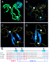
Fig. 3. The KAP-1 PHD structure. (A) α-carbon overlay of 10 KAP-1 PHD domain structures. The residues N-terminal to I627 and C-terminal to H670 are disordered and not shown. The average position of the two zinc atoms is represented by white spheres. The blue portion of the ensemble represents the flexible hinge region described in the text. Structural statistics are given in Table I. (B) All-atom view of KAP-1 PHD in the same orientation as in (A). The main chain is colored in gray. Side chains are colored as follows: hydrophobic in yellow, polar (non-charged) in cyan, polar (acidic) in red, and polar (basic) in blue. (C and D) Ribbon diagram of the KAP-1 PHD domain. The β-strands are shown as arrows and zinc ligands in green. (E) Sequence alignment of PHD domains and a schematic depicting zinc ligation.
None
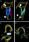
Fig. 4. Comparison of the KAP-1 PHD, PML RING, IEEHV RING and RAG1 RING. Superposition of KAP-1 PHD (with blue β-strands, aa 627–652) and PML RING (magenta β-strands, aa 56–81) (A) and RAG1 RING (yellow β-strands, aa 292–317) (B) from the first metal ligand to the sixth metal ligand. The white spheres represent zinc atoms with the upper zinc atom being site I. The orientation of KAP-1 PHD is the same in both panels. (C) Superposition of zinc-binding site I for KAP-1 (627–632, 647–652), PML (56–71, 76–81), IEEHV (7–12, 28–33) and RAG1 (292–297, 312–317). The metal ligands are colored according to protein: KAP-1 in blue, PML in magenta, IEEHV in green and RAG1 in yellow. (D) Superposition of the conserved hydrophobic core residues N-terminal to metal ligand 5 and C-terminal to metal ligand 6 (L76 and L81 in PML, F28 and I33 in IEEHV, and F312, I317, F647 and H652 in PHD). The side-chains are colored as in (C). The core residues from PHD are noted for clarity. The conserved tryptophan within the PHD family (W664 in KAP-1) is seen here inserting between the other core residues, repositioning the core.
None
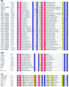
Fig. 5. Amino acid sequence alignment of the PHD, RING and LIM families. Site I metal ligands are colored in magenta and site II metal ligands in blue. Conserved hydrophobic core residues are colored in yellow. DDBJ/EMBL/GenBank accession Nos and PDB codes: KAP-1 (U78773), TIF1α (AAD17258), Mi2α (Q14839), ATRX (P46100), DNMt3A (BAA95556), ING1 (AAG02578), YNJ7_YEAST (P50947), RBB2_HUMAN (P29375), YM42_YEAST (Q03214), YMW5_YEAST (Q04779), YA27_SCHPO (Q09698), YAC5_SCHPO (Q09819), AIRE_HUMAN (O43918), CHD4_HUMAN (Q14839), X169_HUMAN (CAA89909), HT31_ARATH (Q04996), PRH_ARATH (P48785), CHD3_CAEEL (Q22516), CHD3_HUMAN (Q12873), YJ89_YEAST (P47156), FALZ_HUMAN (Q12830), YANC_SCHPO (Q10077), YGN1_YEAST (P53127), YAJ8_SCHPO (Q09908), AF17_HUMAN (Q09908), YGN1_YEAST (P53127), HRX_HUMAN (Q03164), PML (1bor), IEEHV (1chc), RAG1 (1RMD), BRCA1 (A58881), BARD1 (NP_00456), c-Cbl (P22681), MDM2 (CAA41684), MDMX (O15151), MAT (S60157), Z (P18541), Mel-18 (P35227), Crp1LIM1 and LIM2 (1B8T), CRIPrat (1IML), Lin-11 (CAA38240), ISL-1 (CAA3749), MEC-3 (S28390), LMX-1 (B46233).
None
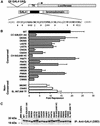
Fig. 6. Mutations in the PHD domain of KAP-1 significantly impair the intrinsic repression activity of KAP-1. (A) Schematic diagram illustrating the reporter plasmid (5×-GAL4-UAS-TK-luciferase) and effector plasmid (GAL4-KAP-1619–835). The primary amino acid sequence of the minimal KAP-1 PHD domain is illustrated. #, conserved amino acids that were mutated. $, non-conserved amino acids in KAP-1 that were mutated to match the corresponding amino acids in TIF1α/TIF1γ. Each mutation was made in the context of a GAL4-KAP-1 (619–835) expression construct. (B) Mutations in the KAP-1 PHD domain disrupt its role in transcriptional repression. All experiments were performed in NIH 3T3 cells with 5 µgof the indicated GAL4 fusion protein and 1 µg of a 5×-GAL4(UAS)-TK-luciferase reporter. A black bar represents wild type, gray bars represent mutations of conserved amino acid residues, and white bars represent substitutions at non-conserved amino acids. (C) Stable expression of each protein was determined via transfection into COS-1 cells followed by immunoprecipitation of [35S]methionine-labeled whole cell extracts with anti-GAL4 (DBD) antiserum (1 µg).
None
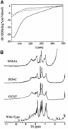
Fig. 7. Some mutations in the PHD domain of KAP-1 disrupt proper folding. (A) Comparison of CD spectra of wild-type KAP-1 PHD (solid line), W664A mutant (dashed/dotted line), P664C mutant (dotted line) and C651F mutant (dashed line) taken at pH 7.5, 25°C. (B) 1D 1H NMR spectra of wild-type and mutant KAP-1 PHD taken at pH 7.5, 30°C. The spectral regions containing the amide and aromatic proton resonances are shown.

References

    1. Aapola U. et al. (2000) Isolation and initial characterization of a novel zinc finger gene, DNMT3L, on 21q22.3, related to the cytosine-5-methyltransferase 3 gene family. Genomics, 65, 293–298. - "VSports在线直播" PubMed
    1. Aasland R., Gibson,T.J. and Stewart,A.F. (1995) The PHD finger: implications for chromatin-mediated transcriptional regulation. Trends Biochem. Sci., 20, 56–59. - PubMed
    1. Barlow P.N., Luisi,B., Milner,A., Elliott,M. and Everett,R. (1994) Structure of the C3HC4 domain by 1H-nuclear magnetic resonance spectroscopy. A new structural class of zinc-finger. J. Mol. Biol., 237, 201–211. - PubMed
    1. Bellon S.F., Rodgers,K.K., Schatz,D.G., Coleman,J.E. and Steitz,T.A. (1997) Crystal structure of the RAG1 dimerization domain reveals multiple zinc-binding motifs including a novel zinc binuclear cluster. Nature Struct. Biol., 4, 586–591. - PubMed
    1. Bochar D.A., Savard,J., Wang,W., Lafleur,D.W., Moore,P., Cote,J. and Shiekhattar,R. (2000) A family of chromatin remodeling factors related to Williams syndrome transcription factor. Proc. Natl Acad. Sci. USA, 97, 1038–1043. - "V体育官网" PMC - PubMed

"VSports app下载" Publication types

MeSH terms

V体育官网入口 - LinkOut - more resources