Skip to main page content
U.S. flag

An official website of the United States government

Dot gov

The . gov means it’s official VSports app下载. Federal government websites often end in . gov or . mil. Before sharing sensitive information, make sure you’re on a federal government site. .

Https

The site is secure V体育官网. The https:// ensures that you are connecting to the official website and that any information you provide is encrypted and transmitted securely. .

. 2000 Nov 20;192(10):1441-52.
doi: 10.1084/jem.192.10.1441.

Mast cells control neutrophil recruitment during T cell-mediated delayed-type hypersensitivity reactions through tumor necrosis factor and macrophage inflammatory protein 2 (VSports最新版本)

Affiliations

"V体育官网" Mast cells control neutrophil recruitment during T cell-mediated delayed-type hypersensitivity reactions through tumor necrosis factor and macrophage inflammatory protein 2

T Biedermann et al. J Exp Med. .

Abstract

Polymorphonuclear leukocytes (PMNs) characterize the pathology of T cell-mediated autoimmune diseases and delayed-type hypersensitivity reactions (DTHRs) in the skin, joints, and gut, but are absent in T cell-mediated autoimmune diseases of the brain or pancreas. All of these reactions are mediated by interferon gamma-producing type 1 T cells and produce a similar pattern of cytokines. Thus, the cells and mediators responsible for the PMN recruitment into skin, joints, or gut during DTHRs remain unknown. Analyzing hapten-induced DTHRs of the skin, we found that mast cells determine the T cell-dependent PMN recruitment through two mediators, tumor necrosis factor (TNF) and the CXC chemokine macrophage inflammatory protein 2 (MIP-2), the functional analogue of human interleukin 8. Extractable MIP-2 protein was abundant during DTHRs in and around mast cells of wild-type (WT) mice but absent in mast cell-deficient WBB6F(1)-Kit(W)/Kit(W-)(v) (Kit(W)/Kit(W)(-v)) mice. T cell-dependent PMN recruitment was reduced >60% by anti-MIP-2 antibodies and >80% in mast cell-deficient Kit(W)/Kit(W)(-v) mice. Mast cells from WT mice efficiently restored DTHRs and MIP-2-dependent PMN recruitment in Kit(W)/Kit(W)-(v) mice, whereas mast cells from TNF(-/)- mice did not VSports手机版. Thus, mast cell-derived TNF and MIP-2 ultimately determine the pattern of infiltrating cells during T cell-mediated DTHRs. .


VSports最新版本 - Figures

Figure 1
Figure 1
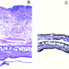
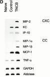
Selective induction of MIP-2 during T cell–dependent CHSRs. TNCB-sensitized mice were challenged with TNCB, 2 d after T cell depletion with anti-Thy 1.2 mAb or sham treatment (A). TNCB-sensitized (B) or naive (C) mice were challenged with TNCB to elicit either a CHSR (B) or an irritant reaction (C). Ear sections were hematoxylin and eosin–stained for microphotographs. mRNA was extracted from ear tissue of untreated mice (left lane), of naive mice challenged for 24 h with TNCB (irritant reaction, middle lane), or of sensitized mice (CHSR, right lane) 24 h after challenge with TNCB (D). MIP-2 was determined by ELISA in protein extracts from ear tissue (E) either from untreated mice (top) or 24 h after challenging either naive mice (irritant reaction, middle) or sensitized mice with TNCB (CHSR, bottom). Hapten treatment, protein and mRNA extractions, reverse transcription, amplification, and evaluation were performed as described in Materials and Methods. DL, detection limit; MCP-1, monocyte chemoattractant protein 1.
Figure 1
Figure 1
Selective induction of MIP-2 during T cell–dependent CHSRs. TNCB-sensitized mice were challenged with TNCB, 2 d after T cell depletion with anti-Thy 1.2 mAb or sham treatment (A). TNCB-sensitized (B) or naive (C) mice were challenged with TNCB to elicit either a CHSR (B) or an irritant reaction (C). Ear sections were hematoxylin and eosin–stained for microphotographs. mRNA was extracted from ear tissue of untreated mice (left lane), of naive mice challenged for 24 h with TNCB (irritant reaction, middle lane), or of sensitized mice (CHSR, right lane) 24 h after challenge with TNCB (D). MIP-2 was determined by ELISA in protein extracts from ear tissue (E) either from untreated mice (top) or 24 h after challenging either naive mice (irritant reaction, middle) or sensitized mice with TNCB (CHSR, bottom). Hapten treatment, protein and mRNA extractions, reverse transcription, amplification, and evaluation were performed as described in Materials and Methods. DL, detection limit; MCP-1, monocyte chemoattractant protein 1.
Figure 2
Figure 2
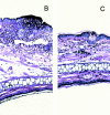
A critical role of MIP-2 for PMN recruitment during CHSRs. TNCB-sensitized mice were challenged with hapten 1.5 h after intracutaneous injection of either preimmune serum or anti–MIP-2 serum (A). Representative microphotographs of hematoxylin and eosin–stained CHSR reactions in mice pretreated with preimmune serum (B) or anti–MIP-2 serum (C). MPO activity in protein extracts from ear tissues (D) challenged with TNCB after pretreatment with either preimmune serum or anti–MIP-2 serum
Figure 2
Figure 2
A critical role of MIP-2 for PMN recruitment during CHSRs. TNCB-sensitized mice were challenged with hapten 1.5 h after intracutaneous injection of either preimmune serum or anti–MIP-2 serum (A). Representative microphotographs of hematoxylin and eosin–stained CHSR reactions in mice pretreated with preimmune serum (B) or anti–MIP-2 serum (C). MPO activity in protein extracts from ear tissues (D) challenged with TNCB after pretreatment with either preimmune serum or anti–MIP-2 serum
Figure 3
Figure 3
MIP-2 production by mast cells in vivo and in vitro. Microphotograph of an ear section stained for mast cells (A) with alcian blue and with anti–MIP-2 serum using immunoperoxidase staining (arrows). Controls using only primary or secondary Ab or preimmune serum showed no significant staining. MIP-2 mRNA expression of cultured mast cells (B) 24 h after stimulation with medium, ionomycin, or ionomycin and IL-1. Reverse transcription PCR was carried out with primers for MIP-2 mRNA (30 cycles). IL-1 alone did not induce MIP-2 mRNA expression. MIP-2 content in the supernatants from mast cells stimulated for 24 h with ionomycin and IL-1 was determined by ELISA (C). No or only very minor MIP-2 mRNA or MIP-2 protein was detected when mast cells were stimulated with either ionomycin or IL-1 alone.
Figure 3
Figure 3
MIP-2 production by mast cells in vivo and in vitro. Microphotograph of an ear section stained for mast cells (A) with alcian blue and with anti–MIP-2 serum using immunoperoxidase staining (arrows). Controls using only primary or secondary Ab or preimmune serum showed no significant staining. MIP-2 mRNA expression of cultured mast cells (B) 24 h after stimulation with medium, ionomycin, or ionomycin and IL-1. Reverse transcription PCR was carried out with primers for MIP-2 mRNA (30 cycles). IL-1 alone did not induce MIP-2 mRNA expression. MIP-2 content in the supernatants from mast cells stimulated for 24 h with ionomycin and IL-1 was determined by ELISA (C). No or only very minor MIP-2 mRNA or MIP-2 protein was detected when mast cells were stimulated with either ionomycin or IL-1 alone.
Figure 4
Figure 4
Mast cell–derived MIP-2 is required for PMN recruitment during CHSRs. Mast cell (MC)-deficient KitW/Kit W-v mice, congenic WT mice, or mast cell–reconstituted KitW/KitW-v mice were sensitized and challenged with TNCB, 1.5 h after pretreatment with either anti–MIP-2 or preimmune serum. Specific increase of ear thickness 24 h after the challenge (A). Microphotographs of hematoxylin and eosin–stained ear sections of CHSR reaction in a WT (B), KitW/KitW -v (C), and mast cell–reconstituted KitW/KitW -v mouse (D). MPO activity was determined in protein extracts from ear tissue of the indicated four experimental groups (E). T cells from unsensitized WT or from draining lymph nodes of sensitized KitW/KitW -v, congenic WT, or mast cell–reconstituted KitW/KitW -v mice were stimulated for 72 h with either naive or TNBS-modified APCs; [3H]thymidine was added for the final 6 h (F). MIP-2 protein in ear tissue was determined from the three experimental groups, 24 h after the TNCB challenge (G). Asterisks indicate significant differences in ear thickness between groups of animals (P < 0.05). DL, detection limit.
Figure 4
Figure 4
Mast cell–derived MIP-2 is required for PMN recruitment during CHSRs. Mast cell (MC)-deficient KitW/Kit W-v mice, congenic WT mice, or mast cell–reconstituted KitW/KitW-v mice were sensitized and challenged with TNCB, 1.5 h after pretreatment with either anti–MIP-2 or preimmune serum. Specific increase of ear thickness 24 h after the challenge (A). Microphotographs of hematoxylin and eosin–stained ear sections of CHSR reaction in a WT (B), KitW/KitW -v (C), and mast cell–reconstituted KitW/KitW -v mouse (D). MPO activity was determined in protein extracts from ear tissue of the indicated four experimental groups (E). T cells from unsensitized WT or from draining lymph nodes of sensitized KitW/KitW -v, congenic WT, or mast cell–reconstituted KitW/KitW -v mice were stimulated for 72 h with either naive or TNBS-modified APCs; [3H]thymidine was added for the final 6 h (F). MIP-2 protein in ear tissue was determined from the three experimental groups, 24 h after the TNCB challenge (G). Asterisks indicate significant differences in ear thickness between groups of animals (P < 0.05). DL, detection limit.
Figure 5
Figure 5
CHSRs in KitW/KitW -v mice depend on mast cell–derived TNF. Kit W/KitW -v mice were reconstituted with mast cells (MC) from either congenic WT or TNF−/− mice, sensitized, and challenged with TNCB as described in Materials and Methods. The increase of ear thickness was determined 24 h after the challenge (A). The asterisk indicates significant difference in ear thickness (P < 0.05). (B) Mast cells from TNF−/− mice in ear tissue from KitW/KitW -v mice stained with toluidine blue (arrowheads).
Figure 5
Figure 5
CHSRs in KitW/KitW -v mice depend on mast cell–derived TNF. Kit W/KitW -v mice were reconstituted with mast cells (MC) from either congenic WT or TNF−/− mice, sensitized, and challenged with TNCB as described in Materials and Methods. The increase of ear thickness was determined 24 h after the challenge (A). The asterisk indicates significant difference in ear thickness (P < 0.05). (B) Mast cells from TNF−/− mice in ear tissue from KitW/KitW -v mice stained with toluidine blue (arrowheads).
Figure 6
Figure 6
MIP-2 immunoreactivity in tissue mast cells from TNF−/− mice. KitW/KitW -v mice were reconstituted with mast cells from either TNF−/− mice (A and B) or congenic WT mice (C). These animals and, in addition, TNF−/− mice (D), were sensitized and challenged with TNCB. MIP-2 (A, C, and D) immunoreactivity was analyzed in fixed cryosections as described in Materials and Methods. (B) Analysis of immunoreactivity in the absence of the primary Ab. Similarly, no staining was found in the corresponding controls for C and D.

References

    1. Burmester G.R., Daser A., Kamradt T., Krause A., Mitchison N.A., Sieper J., Wolf N. Immunology of reactive arthritides. Annu. Rev. Immunol. 1995;13:229–250. - V体育官网入口 - PubMed
    1. Feldmann M., Brennan F.M., Maini R.N. Role of cytokines in rheumatoid arthritis. Annu. Rev. Immunol. 1996;14:397–440. - PubMed (VSports最新版本)
    1. Edwards S.W., Hallett M.B. Seeing the wood for the treesthe forgotten role of neutrophils in rheumatoid arthritis. Immunol. Today. 1997;18:320–324. - PubMed
    1. Strober W., Ehrhardt R.O. Chronic intestinal inflammationan unexpected outcome in cytokine or T cell receptor mutant mice. Cell. 1993;75:203–205. - PubMed
    1. Romagnani S. Th1/Th2 cells. Inflamm. Bowel Dis. 1999;5:285–294. - V体育ios版 - PubMed

Publication types

MeSH terms

V体育平台登录 - Substances