Skip to main page content (V体育平台登录)
U.S. flag

An official website of the United States government

Dot gov

The . gov means it’s official VSports app下载. Federal government websites often end in . gov or . mil. Before sharing sensitive information, make sure you’re on a federal government site. .

Https

The site is secure. The https:// ensures that you are connecting to the official website and that any information you provide is encrypted and transmitted securely V体育官网. .

. 2000 Dec;68(12):6542-53.
doi: 10.1128/IAI.68.12.6542-6553.2000.

V体育平台登录 - Identification and characterization of the scl gene encoding a group A Streptococcus extracellular protein virulence factor with similarity to human collagen

Affiliations

Identification and characterization of the scl gene encoding a group A Streptococcus extracellular protein virulence factor with similarity to human collagen

S Lukomski et al. Infect Immun. 2000 Dec.

Abstract

Group A Streptococcus (GAS) expresses cell surface proteins that mediate important biological functions such as resistance to phagocytosis, adherence to plasma and extracellular matrix proteins, and degradation of host proteins VSports手机版. An open reading frame encoding a protein of 348 amino acid residues was identified by analysis of the genome sequence available for a serotype M1 strain. The protein has an LPATGE sequence located near the carboxy terminus that matches the consensus sequence (LPXTGX) present in many gram-positive cell wall-anchored molecules. Importantly, the central region of this protein contains 50 contiguous Gly-X-X triplet amino acid motifs characteristic of the structure of human collagen. The structural gene (designated scl for streptococcal collagen-like) was present in all 50 GAS isolates tested, which together express 21 different M protein types and represent the breadth of genomic diversity in the species. DNA sequence analysis of the gene in these 50 isolates found that the number of contiguous Gly-X-X motifs ranged from 14 in serotype M6 isolates to 62 in a serotype M41 organism. M1 and M18 organisms had the identical allele, which indicates very recent horizontal gene transfer. The gene was transcribed abundantly in the logarithmic but not stationary phase of growth, a result consistent with the occurrence of a DNA sequence with substantial homology with a consensus Mga binding site immediately upstream of the scl open reading frame. Two isogenic mutant M1 strains created by nonpolar mutagenesis of the scl structural gene were not attenuated for mouse virulence as assessed by intraperitoneal inoculation. In contrast, the isogenic mutant derivative made from the M1 strain representative of the subclone most frequently causing human infections was significantly less virulent when inoculated subcutaneously into mice. In addition, both isogenic mutant strains had significantly reduced adherence to human A549 epithelial cells grown in culture. These studies identify a new extracellular GAS virulence factor that is widely distributed in the species and participates in adherence to host cells and soft tissue pathology. .


Figures
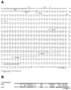
FIG. 1
FIG. 1
Nucleotide and amino acid sequence of the scl gene and Scl protein in serotype M1 GAS. (A) The scl coding sequence consists of 1,047 bp (nucleotides 77 to 1123) and encodes a polypeptide of 348 amino acids. The presumed scl promoter region contains a predicted ribosome-binding site (RBS) and −10 and −35 regions. A predicted transcription site, +1, is denoted by a dot. A potential binding site for the streptococcal positive transcription regulator, Mga, overlapping with the −35 region is shown. A potential transcription terminator (tt) consisting of two inverted repeats was identified downstream of scl gene. The predicted ATG start codon and the TAA stop codon are shown in boldface type. SS, signal sequence showing the predicted cleavage site (arrowhead); V, V region of the mature Scl protein; CL, CL region consisting of 50 GXX triplets (boxed); L, L region with PGEKAPEKS repeats (underlined); WM, cell-associated Scl part containing a cell wall segment with the LPATGE motif (shaded), and cell membrane hydrophobic region with a charged tail. (B) Alignment of the consensus Mga binding site and putative promoter region of the scl gene. The consensus Mga binding site was identified on the basis of sequence alignments of several Mga-regulated genes (34). The essential bases are shown in boldface type, and essential sequences within the potential Mga binding site of the scl gene promoter which do not match with the consensus sequence are shown in lowercase type. Twenty-two of 29 (76%) essential bases are identical. Underlined bases in the putative promoter region of the scl gene promoter denote a potential −35 promoter region.
FIG. 2
FIG. 2
scl gene expression in M1 GAS strains. (A) Northern blot analysis of the total RNA isolated from cultures of MGAS 6708 and MGAS 5005 harvested during the logarithmic (EL [early logarithmic] [OD600 ≈ 0.5]) or stationary (ES [early stationary] [OD600 ≈ 0.8] or LS [late stationary, overnight]) phase of growth. RNA (15 μg) was loaded in each lane. A DNA probe consisting of the entire scl gene was amplified by PCR from the MGAS 6708 source strain and labeled by biotinylation. The blot was developed with streptavidin-peroxidase conjugate and a chemiluminescence detection method was used to visualize hybridizing bands. A single transcript of ∼1.2 kb was identified in log-phase cultures. scl gene transcription decreased markedly during the stationary phase of growth. RNA size markers (in kilobases) were used to estimate the size of the scl transcript. (B) Western blot analysis of the cell wall-associated protein fractions from exponential GAS cultures. Both wild-type (wt) M1 serotype MGAS 6708 and MGAS 5005 strains have an immunoreactive cell wall-associated protein migrating at ∼49 kDa. Molecular mass standards (in kilodaltons) are shown.
FIG. 3
FIG. 3
Size variation in the scl gene. The complete scl gene and putative promoter region were amplified by PCR and analyzed by agarose gel electrophoresis. All 50 strains tested yielded a single PCR product, and representative examples of the size variants identified are shown. (A) Size variation in the scl gene in different M-type GAS strains. (B) scl gene size variation among strains of certain M types, including M12, M28, and M56.
FIG. 4
FIG. 4
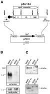
scl gene expression in wild-type and mutant M1 GAS. (A) Schematic representation of suicide plasmid pSL134 used for nonpolar inactivation of the scl gene in serotype M1 GAS. Most of the scl gene sequence (969 of 1,047 bp) was replaced with a nonpolar spectinomycin resistance cassette (spc2) inserted in frame. To enhance the likelihood of homologous recombination, ∼400 bp of DNA upstream and downstream of the scl ORF also was cloned. pSL134 is a suicide plasmid that cannot replicate in GAS. Ss, signal sequence; V, variable region; CL, collagen-like region; WM, cell wall and cell membrane region; tt, transcription terminator; MCS, multiple cloning site. (B) Northern blot analysis of the total RNA from logarithmic-phase GAS cultures. Samples (15 μg) were blotted and hybridized with scl- and recA-specific biotinylated DNA probes and detected by chemiluminescence. The scl-specific transcript is detected in the wild-type (wt) but not mutant sample. Control hybridization with the recA probe showed a positive signal for both the wild-type and scl mutants. Identical results were obtained for the MGAS 6708 isogenic pair (data not shown). (C) Western blot analysis of the cell wall-associated protein fractions obtained from the wild-type and scl mutant strains. Scl is detected in the samples prepared from the wild-type but not mutant strains.
FIG. 5
FIG. 5
Kaplan-Meier survival curves showing mouse lethality caused by the wild type (open symbols) and isogenic scl-inactivated mutants (closed symbols). Experiments were performed with CD-1 Swiss mice inoculated i.p. with M1 type GAS. (A) Determination of the inoculum size for MGAS 6708 wild-type GAS. Inocula used were 1.1 × 10⁹ CFU (circles), 7.5 × 10⁸ CFU (diamonds), and 3.3 × 10⁸ CFU or less (triangle). (B) MGAS 6708 wild-type strain (1.1 × 10⁹ CFU [n = 15 mice/group]) and MGAS 6708 mutant (1.2 × 10⁹ CFU [n = 15 mice/group]). (C) MGAS 5005 wild-type (2.2 × 10⁷ CFU [n = 17 mice/group]) and MGAS 5005 mutant (2.3 × 10⁷ CFU [n = 18 mice/group]). The differences in mouse mortality between the wild-type and mutant strains were not statistically significant.
FIG. 6
FIG. 6
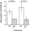
Adherence of isogenic M1 GAS strains to cultured human epithelial cells. Mid-log phase cultures of the wild-type strains (open bars) and scl-inactivated mutants (filled bars) were incubated with A549 epithelial cells for 1 h. Unattached bacteria were removed by washing. The attached GAS were stained and counted microscopically. The results are presented as mean numbers of GAS cells attached to each A549 cell ± standard error of the mean (error bars). The difference in adherence between the wild type and mutant derivatives was significant by t test for both MGAS 5005 and MGAS 6708 strain pairs.
FIG. 7
FIG. 7
Schematic representation of variation in the Scl protein sequence among three different M-type GAS strains. The amino acid sequence of the Scl protein from M1 (source strain), M6, and M28 (scl pattern I [Table 1]) GAS strains is shown. The length of each Scl domain among all 50 GAS strains tested is shown as the number of amino acid residues. Glycine residues within the CL region are shown in boldface type. Polymorphic sites within the signal sequence and linker region are in boldface type and italicized. The signal sequence consists of 37 amino acids in all strains tested. In addition to the G31V substitution in the Scl signal sequence of the M6 strain, the following amino acid replacements were found in other GAS strains: H7Y, L9I, L12Q, and T19I. The alanine (A256 in M1 strain) residue (shown in lowercase type) located after the last GEK triplet motif is counted as the first residue of the linker region. The cell wall region contains the charged-tail sequence KRKENN (underlined). The KRKEEN motif in the M6 and M28 strains is shown in brackets because DNA sequence analysis did not identify all of the gene due to primer placement. The following polymorphic changes were identified in the cell wall region in all strains tested (amino acid residue numbers correspond to the sequence in the M1 strain): N292S and T335I.

"VSports手机版" References

    1. Akesson P, Sjoholm A G, Bjorck L. Protein SIC, a novel extracellular protein of Streptococcus pyogenes interfering with complement function. J Biol Chem. 1996;271:1081–1088. - PubMed
    1. Ashbaugh C D, Warren H B, Carey V J, Wessels M R. Molecular analysis of the role of the group A streptococcal cysteine protease, hyaluronic acid capsule, and M protein in a murine model of human invasive soft-tissue infection. J Clin Investig. 1998;102:550–560. - PMC - PubMed
    1. Camper L, Hellman U, Lundgren-Akerlund E. Isolation, cloning, and sequence analysis of the integrin subunit α10, a β1-associated collagen binding integrin expressed on chondrocytes. J Biol Chem. 1998;273:20383–20389. - PubMed
    1. Caparon M G, Scott J R. Identification of a gene that regulates expression of M protein, the major virulence determinant of group A streptococci. Proc Natl Acad Sci USA. 1987;84:8677–8681. - "VSports app下载" PMC - PubMed
    1. Caparon M G, Stephens D S, Olsen A, Scott J R. Role of M protein in adherence of group A streptococci. Infect Immun. 1991;59:1811–1817. - PMC - PubMed

"V体育2025版" Publication types

V体育官网入口 - MeSH terms

LinkOut - more resources