<small lang="sq97gUX"></small> "V体育平台登录" Skip to main page content
U.S. flag

An official website of the United States government

Dot gov

The . gov means it’s official. Federal government websites often end in . gov or . mil. Before sharing sensitive information, make sure you’re on a federal government site VSports app下载. .

Https

The site is secure. The https:// ensures that you are connecting to the official website and that any information you provide is encrypted and transmitted securely. V体育官网.

. 2000 Nov 7;97(23):12869-74.
doi: 10.1073/pnas.220301797.

Functional mosaic organization of mouse olfactory receptor neurons (V体育平台登录)

Affiliations

"VSports" Functional mosaic organization of mouse olfactory receptor neurons

M Ma et al. Proc Natl Acad Sci U S A. .

V体育ios版 - Abstract

In contrast to rapid progress in the molecular biology of olfaction, there are few physiological data to characterize the odor response properties of different populations of olfactory receptor neurons (ORNs) and their spatial distributions across the epithelium, which is essential for understanding the coding mechanisms underlying odor discrimination and recognition. We have tested the hypothesis that the ORNs are arranged in a functional mosaic, using an intact epithelial preparation from the mouse, in which odor responses of many ORNs in situ can be monitored simultaneously with calcium imaging techniques. ORNs responding to a given odor were widely distributed across epithelium and intermingled with ORNs responding to other odors. Tight clusters of ORNs responding to the same odor were observed. For a given odor, more ORNs were recruited when the concentration was increased. ORNs were able to distinguish between pairs of enantiomers by showing distinct but somewhat overlapping patterns VSports手机版. The results provide evidence regarding the response spectra of ORNs in situ, supporting the combinatorial coding of odor quality and intensity by different ORN subsets. .


"V体育平台登录" Figures
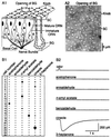
Figure 1
Figure 1
(A) Visualization of dendritic knobs of mouse ORNs. (A1) Schematic drawing of olfactory epithelium. BG, Bowman's gland. SC, supporting cell. The 10–15 cilia emanating from each knob are not drawn. (A2) The dendritic knobs of mouse ORNs were seen as bright spots in an en face view under infrared differential interference contrast microscope. (B) Responses to different odors by single ORNs, revealed by perforated patch-clamp recordings. (B1) The table summarizes the responses of 25 cells to six different odors. Identical 100-ms pulses of six different odors at 300 μM were sequentially delivered to a cell. The interval between two consecutive pulses was >30 sec. If a cell did not respond to a mixture of the six odors, it was considered not to respond to any individual odor. The area of a filled circle represents the relative amplitude of the odor-induced current; a dash indicates no detectable current. The holding potential was −60 mV. The cells were chosen randomly from all areas of olfactory epithelium. (B2) Raw data from cell 22. Of the six odors, the cell responded only to cineole.
Figure 2
Figure 2
Imaging odor responses of mouse ORNs in the intact epithelial preparation by calcium imaging techniques. (A) The preparation was viewed under fluorescence illumination, after being loaded with calcium green. (B) A total of 216 dendritic knobs are mapped for further analysis. Each circle represents a cell. Red circles (30 of 216) stand for the responsive cells to a 4-sec odor stimulus (a mixture containing 10 μM of each of the six odors listed in Fig. 1B). (C) The fluorescence intensity changes (ΔF/F) induced by the odor stimulus are measured and calculated for individual dendritic knobs; the traces corresponding to cells a–f are shown. (D) Comparison between transmitted (Left) and fluorescent (Right) images confirmed that the bright spots were dendritic knobs. The arrows mark the dendritic knobs. SC, supporting cell. (E) The fluorescence changes (ΔF/F) induced by odor stimulation was caused by calcium entry. Perfusion of saline (trace 1) or removal of Ca2+ (trace 3) eliminated odor-induced fluorescence changes. The stimulus was saline (trace 1) or an odor mixture as in Fig. 1B (traces 2–4). The bath was normal (traces 1, 2, and 4) or low Ca2+ saline (trace 3).
Figure 3
Figure 3
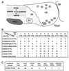
Summary of the data obtained from the septal epithelium. (A) The positions of the recorded areas (rectangles of 215 μm by 172 μm) are numbered from 1 to 12. The standard contour of the epithelium was obtained from a 6-week-old female mouse. RE, respiratory epithelium. OE, olfactory epithelium. VNO, vomeronasal organ. SO, septal organ. OB, olfactory bulb. (B) Summary of the number of ORNs in response to different odors. The viewing areas nos. 1–12 shown in A were from 12 different preparations. N (in parentheses) stands for the total number of cells tested in each area. The percentage after each odor is the averaged percentage of cells that responded to the odor from the 12 preparations. A dash indicates the fact that an odor was not tested. All odors were at 50 μM. (C) Summary of the number of ORNs in response to odors with different functional groups. A total of 102 responsive cells were from 1,846 tested. The odors used were alcohols (OH), aldehydes (CHO), and carboxylic acids (COOH) with straight seven or eight carbon chains at a concentration of 50 μM.
Figure 4
Figure 4
Mosaic organization of mouse ORNs in responding to different odors. (A) The preparation was viewed under fluorescence illumination, after being loaded with calcium green. (B) ORNs responded to different odors with distinct patterns. A total of 163 cells are mapped. Responsive ORNs to different odors are represented by different colors. Cells responding to more than one odor are drawn in multiple colors. The numbers in parentheses are the numbers of responsive cells to the given odor(s). Filled and hollow arrows mark the tight clusters of octanal and n-amyl acetate responsive cells, respectively. All odors were at 50 μM.
Figure 5
Figure 5
Mouse olfactory epithelia respond to stronger stimuli by recruiting more ORNs. (A) More cells responded to 50 μM (pink) n-amyl acetate than to 1 μM (red). Of 118 cells, 19 responded to 50 μM and only six responded to 1 μM n-amyl acetate. (B) Cells responding to the other four odors are mapped in the same preparation as shown in A. Responsive ORNs to different odors are represented by different colors. Cells responding to more than one odor are drawn in multiple colors. The numbers in parentheses are the numbers of responsive cells to the given odor(s). All odors were at 50 μM.
Figure 6
Figure 6
Mouse ORNs can distinguish between pairs of enantiomers. ORNs responded to (+) and (−) carvone at different concentrations with distinct patterns. A total of 170 cells are mapped. Responsive ORNs to the two isomers at different concentrations are represented by different colors. The numbers in parentheses are the numbers of responsive cells to the given odor(s).

References

    1. Buck L, Axel R. Cell. 1991;65:175–187. - PubMed
    1. Ressler K J, Sullivan S L, Buck L B. Cell. 1993;73:597–609. - "V体育平台登录" PubMed
    1. Vassar R, Ngai J, Axel R. Cell. 1993;74:309–318. - PubMed
    1. Chess A, Simon I, Cedar H, Axel R. Cell. 1994;78:823–834. - PubMed
    1. Malnic B, Hirono J, Sato T, Buck L B. Cell. 1999;96:713–723. - V体育平台登录 - PubMed

Publication types

Substances

VSports - LinkOut - more resources