"V体育ios版" Skip to main page content
U.S. flag

An official website of the United States government

Dot gov

The . gov means it’s official. Federal government websites often end in . gov or VSports app下载. mil. Before sharing sensitive information, make sure you’re on a federal government site. .

Https

The site is secure. The https:// ensures that you are connecting to the official website and that any information you provide is encrypted and transmitted securely V体育官网. .

. 2000 Sep 1;20(17):6421-30.
doi: 10.1523/JNEUROSCI.20-17-06421.2000.

Passive or active immunization with myelin basic protein promotes recovery from spinal cord contusion (VSports最新版本)

Affiliations

Passive or active immunization with myelin basic protein promotes recovery from spinal cord contusion

E Hauben et al. J Neurosci. .

Erratum in

  • J Neurosci. 2016 Feb 10;36(6):2075

Abstract

Partial injury to the spinal cord can propagate itself, sometimes leading to paralysis attributable to degeneration of initially undamaged neurons. We demonstrated recently that autoimmune T cells directed against the CNS antigen myelin basic protein (MBP) reduce degeneration after optic nerve crush injury in rats. Here we show that not only transfer of T cells but also active immunization with MBP promotes recovery from spinal cord injury. Anesthetized adult Lewis rats subjected to spinal cord contusion at T7 or T9, using the New York University impactor, were injected systemically with anti-MBP T cells at the time of contusion or 1 week later. Another group of rats was immunized, 1 week before contusion, with MBP emulsified in incomplete Freund's adjuvant (IFA). Functional recovery was assessed in a randomized, double-blinded manner, using the open-field behavioral test of Basso, Beattie, and Bresnahan. The functional outcome of contusion at T7 differed from that at T9 (2. 9+/-0. 4, n = 25, compared with 8. 3+/-0. 4, n = 12; p<0. 003). In both cases, a single T cell treatment resulted in significantly better recovery than that observed in control rats treated with T cells directed against the nonself antigen ovalbumin. Delayed treatment with T cells (1 week after contusion) resulted in significantly better recovery (7. 0+/-1; n = 6) than that observed in control rats treated with PBS (2. 0+/-0. 8; n = 6; p<0. 01; nonparametric ANOVA). Rats immunized with MBP obtained a recovery score of 6. 1+/-0. 8 (n = 6) compared with a score of 3. 0+/-0. 8 (n = 5; p<0. 05) in control rats injected with PBS in IFA. Morphometric analysis, immunohistochemical staining, and diffusion anisotropy magnetic resonance imaging showed that the behavioral outcome was correlated with tissue preservation. The results suggest that T cell-mediated immune activity, achieved by either adoptive transfer or active immunization, enhances recovery from spinal cord injury by conferring effective neuroprotection VSports手机版. The autoimmune T cells, once reactivated at the lesion site through recognition of their specific antigen, are a potential source of various protective factors whose production is locally regulated. .


"VSports最新版本" Figures

Fig. 1.
Fig. 1.
Spontaneous recovery from spinal cord contusion at T7 and T9. Rats were subjected to spinal cord contusion under deep anesthesia and immediately injected systemically with PBS. Recovery was assessed by the BBB open-field test at the indicated time points by observers blinded to the treatment received by the rats. Results are expressed as the mean values for each group (error bars indicate SEM). The differences, tested by repeated ANOVA, were significant (T7,n = 25; T9, n = 12;p < 0.003).
Fig. 2.
Fig. 2.
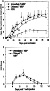
Anti-MBP T cells enhance recovery of voluntary motor activity after spinal cord contusion. A, Twelve rats were deeply anesthetized, laminectomized, and subjected to spinal cord contusion (T9). Six of the rats were then inoculated intraperitoneally with 107 anti-MBP T cells in PBS (black circles), and the rest were injected with PBS (black squares). At the indicated times, locomotor behavior in an open field was scored. The results are expressed as the mean values for each group (error bars indicate SEM). The differences, tested by repeated ANOVA, were significant (p < 0.05). C, In another group of rats, the spinal cords were completely transected and the rats were divided into subgroups receiving either 107anti-MBP T cells or PBS. No significant differences in locomotor behavior were seen between the two subgroups at any time during follow-up. B, D, Course of EAE development in sham-operated rats treated with anti-MBP T cells. Lewis rats were subjected to sham operation (laminectomy but not contusion) and immediately injected with anti-MBP T cells. EAE was evaluated according to a neurological paralysis scale. Values represent means ± SEM.
Fig. 3.
Fig. 3.
Retrograde labeling of cell bodies in the red nucleus. Three months after spinal contusion at the level of T9 followed immediately by immunization with anti-MBP T cells or injection of PBS, three rats from each group were reanesthetized, and the dye rhodamine dextran amine (Fluoro-ruby) was applied below the site of contusion. Sections taken through the red nucleus were inspected and analyzed qualitatively and quantitatively by fluorescence and confocal microscopy. Significantly more labeled red nuclei were seen in the rats treated with anti-MBP T cells than in the PBS-treated rats (p = 0.046; Student's ttest, with correction for thickness and size of neurons). The bar graph shows the average of the total numbers of labeled red nuclei per brain. The behavioral scores of the three rats were 10.5, 12, and 12.75 in the experimental group and 6, 8, and 8.5 in the control group. The bar graph shows the mean ± SD values.
Fig. 4.
Fig. 4.
Spinal cord recovery after delayed administration of anti-MBP T cells. One week after contusion at the level of T7, rats (n = 15) were randomly divided into two groups for injection with either PBS (n = 8) or 107 anti-MBP T cells (n = 7).A, The graph shows the mean ± SEM locomotor activity scores at the indicated periods after T7 contusion. Plateau values reached by the anti-MBP T cell-treated rats were significantly higher than those reached by the controls (p< 0.001; ANOVA). For comparison, a similar experiment using five PBS-treated rats and six rats treated immediately with anti-MBP T cells is also shown here. There was no difference between the immediate and the delayed T cell treatment in terms of the maximal plateau values. Another group of rats with contusion at T7 received anti-OVA T cells. No effect was observed relative to PBS-treated rats. B, The course of EAE development in sham-operated rats treated with anti-MBP T cells. Lewis rats were subjected to sham operation (laminectomy but not contusion) and immediately injected with anti-MBP T cells. EAE was evaluated according to a neurological paralysis scale. Values represent means ± SEM.
Fig. 5.
Fig. 5.
Promotion of spinal cord recovery by active immunization with MBP. Six rats were immunized subcutaneously with MBP in IFA, and six were injected with PBS in IFA. One week later, the rats were deeply anesthetized, laminectomized, and subjected to spinal cord contusion at T7. At the indicated times, locomotor behavior in an open field was scored. The results are expressed as the mean values for each group (error bars indicate SEM). The differences, tested by repeated ANOVA, were significant (p < 0.05).
Fig. 6.
Fig. 6.
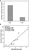
Correlation between BBB locomotor score and the number of retrogradely labeled red nuclei. Rats were actively immunized with MBP, and this was followed 1 week later by contusion. Two months after injury, a dye was applied below the primary contusion site. Five days later, the rats were killed, and their brains were excised and analyzed. A, Bar graphs show the number of labeled rubrospinal neurons in contused rats immunized with MBP in IFA or injected with PBS in IFA. B, Correlation between BBB locomotor score and the number of retrogradely labeled rubrospinal neurons. The graph shows the number of labeled neurons and the BBB score for each rat. The data for all of the examined rats are included.
Fig. 7.
Fig. 7.
Maps showing diffusion anisotropy of the contused spinal cords. Rats were deeply anesthetized, and their excised spinal cords were immediately fixed and placed in 5 mm nuclear magnetic resonance tubes. The figure shows representative maps of spinal cords of anti-MBP T cell-treated rats and control rats, after contusion at T8–T9. Colors correspond to anisotropy ratios. The maps show the preservation of longitudinally ordered tissue at the lesion sites of the anti-MBP T cell-treated rats. Note that, in the controls, the site of injury is much larger than in rats from the anti-MBP T cell-treated group.
Fig. 8.
Fig. 8.
Phase microscopy and histochemical staining of contused spinal cords. Rats were subjected to spinal cord contusion (T7) and were treated immediately with anti-MBP T cells or with PBS. After 5 months, the spinal cords from three PBS-treated controls and three anti-MBP T cell-treated rats were excised and processed for confocal microscopy. Representative micrographs from each group are shown. Note the large gap and the cysts in the neural tissues of a PBS-treated rat (a) compared with an anti-MBP T cell-treated rat (b). For comparison, a phase micrograph of a sham-operated spinal cord is included (c).
Fig. 9.
Fig. 9.
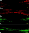
Fluorescence micrographs of spinal cords stained for GFAP and NFs. Sections taken from the preparation described in Figure 8 were analyzed by immunohistochemical staining for GFAP to delineate the site of injury. Note the gap in staining of the PBS-treated cord (a) compared with the anti-MBP T cell-treated cord (b). Sections were also analyzed for NFs. The gap between the severed ends of axons is smaller in the rat treated with anti-MBP T cells (d) than in the PBS-treated rat (c).
Fig. 10.
Fig. 10.
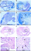
Light microscopy of cross-sections from the site of injury. Transverse sections (4 μm) were taken from the center of the lesion site of contused spinal cords treated with anti-MBP T cells or PBS and stained with H&E (E–H) or luxol (A–D). A, C,E, and G show sections from control rats.B, D, F, andH show sections from rats treated with anti-MBP T cells.Arrowheads and arrows point to myelinated axons and neuronal cell bodies, respectively. C andD are enlargements of the boxed areas seen inA and B; G andH are enlargements of E andF.

"VSports" References

    1. Artis D, Humphreys NE, Bancroft AJ, Rothwell NJ, Potten CS, Grencis RK. Tumor necrosis factor alpha is a critical component of interleukin 13-mediated protective T helper cell type 2 responses during helminth infection. J Exp Med. 1999;190:953–962. - PMC - PubMed
    1. Basso DM, Beattie MS, Bresnahan JC. A sensitive and reliable locomotor rating scale for open field testing in rats. J Neurotrauma. 1995;12:1–21. - PubMed
    1. Basso DM, Beattie MS, Bresnahan JC. Graded histological and locomotor outcomes after spinal cord contusion using the NYU weight-drop device versus transection. Exp Neurol. 1996;139:244–256. - "V体育官网入口" PubMed
    1. Bavetta S, Hamlyn PJ, Burnstock G, Lieberman AR, Anderson PN. The effects of FK506 on dorsal column axons following spinal cord injury in adult rats: neuroprotection and local regeneration. Exp Neurol. 1999;158:382–393. - PubMed
    1. Beattie MS, Bresnahan JC, Komon J, Tovar CA, Van Meter M, Anderson DK, Faden AI, Hsu CY, Noble LJ, Salzman S, Young W. Endogenous repair after spinal cord contusion injuries in the rat. Exp Neurol. 1997;148:453–463. - PubMed

Publication types

VSports注册入口 - Substances