Skip to main page content
U.S. flag

An official website of the United States government

Dot gov

The . gov means it’s official. Federal government websites often end in . gov or . mil. Before sharing sensitive information, make sure you’re on a federal government site VSports app下载. .

Https

The site is secure. The https:// ensures that you are connecting to the official website and that any information you provide is encrypted and transmitted securely. V体育官网.

. 2000 Sep;74(18):8252-61.
doi: 10.1128/jvi.74.18.8252-8261.2000.

Human immunodeficiency virus type 1 Vif protein is an integral component of an mRNP complex of viral RNA and could be involved in the viral RNA folding and packaging process

Affiliations

Human immunodeficiency virus type 1 Vif protein is an integral component of an mRNP complex of viral RNA and could be involved in the viral RNA folding and packaging process

H Zhang et al. J Virol. 2000 Sep.

Abstract

Virion infectivity factor (Vif) is a protein encoded by human immunodeficiency virus types 1 and 2 (HIV-1 and -2) and simian immunodeficiency virus, plus other lentiviruses, and is essential for viral replication either in vivo or in culture for nonpermissive cells such as peripheral blood lymphoid cells, macrophages, and H9 T cells. Defects in the vif gene affect virion morphology and reverse transcription but not the expression of viral components VSports手机版. It has been shown that Vif colocalizes with Gag in cells and Vif binds to the NCp7 domain of Gag in vitro. However, it seems that Vif is not specifically packaged into virions. The molecular mechanism(s) for Vif remains unknown. In this report, we demonstrate that HIV-1 Vif is an RNA-binding protein and specifically binds to HIV-1 genomic RNA in vitro. Further, Vif binds to HIV-1 RNA in the cytoplasm of virus-producing cells to form a 40S mRNP complex. Coimmunoprecipitation and in vivo UV cross-linking assays indicated that Vif directly interact with HIV-1 RNA in the virus-producing cells. Vif-RNA binding could be displaced by Gag-RNA binding, suggesting that Vif protein in the mRNP complex may mediate viral RNA interaction with HIV-1 Gag precursors. Furthermore, we have demonstrated that these Vif mutants that lose the RNA binding activity in vitro do not support vif-deficient HIV-1 replication in H9 T cells, suggesting that the RNA binding capacity of Vif is important for its function. Further studies regarding Vif-RNA interaction in virus-producing cells will be important for studying the function of Vif in the HIV-1 life cycle. .


Figures

FIG. 1
FIG. 1
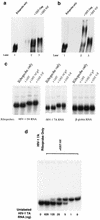
HIV-1 Vif binds to polynucleotide homopolymers in vitro. (a) In vitro-translated, 35S-labeled HIV-1 Vif was mixed with polynucleotide homopolymers [poly(G), poly(A), poly(C), and poly(U)]-conjugated agarose beads, respectively. (b) In vitro-translated, 35S-labeled HIV-1 Vif was mixed with poly(G)-conjugated agarose beads in the presence of NaCl at various concentrations. After a binding and washing procedure, the bead-associated Vif protein was fractionated by SDS-PAGE.
FIG. 2
FIG. 2
Gel shift assay to analyze the interaction between Vif and RNA. (a and b) GST-Gag and GST-Vif (10 pmol) were allowed to bind with the HIV-1 riboprobe 7A (located in the HIV-1 RNA genome at nucleotides 5104 to 5287) in the presence of 750 ng of tRNA (a) or poly(dI-dC) (b). (c) GST and GST-fusion proteins (GST-NCp7 and GST-Vif) (10 pmol) were allowed to bind with the HIV-1 riboprobe 5A (located in the HIV-1 RNA genome at nucleotides 3677 to 3925) and riboprobe 7A, respectively. As a control, a riboprobe generated from the γ-actin gene (nucleotides 81 to 280) was also allowed to bind with GST and GST-Vif. The RNA-protein mixtures were then fractionated on a 5% native TBE gel. (d) GST-Vif protein (10 pmol) were allowed to bind with HIV-1 riboprobe 7A in the presence of in vitro-transcribed, unlabeled HIV-1 7A at various concentrations. The mixtures were then fractionated on a 5% native TBE gel, followed by autoradiography.
FIG. 3
FIG. 3
In vitro UV light-induced cross-linking assay. GST-Vif was mixed with HIV-1 riboprobes 2C (located in the HIV-1 RNA genome at nucleotides 1467 to 1677), 5A, and 7A. As controls, GST and GST-NCp7 were also mixed with HIV-1 riboprobes, while GST-Vif was mixed with an γ-actin riboprobe (lane 10). The mixtures were irradiated with UV light, followed by digestion with RNase A. The samples were then fractionated by SDS-PAGE, and protein-associated 32P-labeled RNA was visualized via autoradiography.
FIG. 4
FIG. 4
Copurification of unspliced HIV-1 RNA with Vif by rate-zonal sedimentation. HIV-1NL4-3-infected H9 cells were lysed, and the postnuclear fraction was divided into two parts; one part was treated with RNase A (1 μg/ml), and the other part was treated with the RNase inhibitor RNasin (320 U/ml). Both portions were then placed onto a 15 to 30% sucrose gradient for ultracentrifugation. Twelve fractions were collected, and Vif protein in all fractions was detected via Western blotting. The sedimentation coefficient was calculated as described previously (44, 70). HIV-1 unspliced and spliced RNAs and γ-globin RNA were detected by RT-PCR.
FIG. 5
FIG. 5
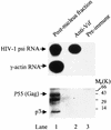
Binding between Vif and HIV-1 RNA analyzed by coimmunoprecipitation assay. Fraction 8 in the -RNase A panel in Fig. 4 was mixed with anti-Vif antibody or preimmune rabbit serum, as well as protein A-conjugated Sepharose beads. The bead-associated HIV-1 unspliced RNA and host cell γ-actin RNA were then detected by RT-PCR, while the bead-associated HIV-1 Gag protein was detected with an anti-p7 antibody via Western blotting.
FIG. 6
FIG. 6
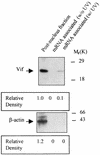
Interaction between Vif and mRNA analyzed by in vivo UV cross-linking assay. HIV-1NL4-3-infected H9 cells were washed and irradiated with UV light. The cells were lysed, and mRNA and associated proteins were then isolated from the postnuclear fraction by oligo(dT)-cellulose chromatography. After digestion with RNase A, the mRNA-associated proteins were detected via Western blotting with either anti-Vif or anti-β-actin antibody.
FIG. 7
FIG. 7
Interaction between Vif, RNA, and Gag in vitro. (a) In vitro-translated, 35S-labeled Vif protein was subjected to binding with GST-Gag-conjugated agarose beads in the presence of free poly(G), HIV-1 7A RNA, or γ-actin RNA. After washing, the bead-associated Vif was fractionated by SDS-PAGE. (b) In vitro-translated, 35S-labeled Vif protein was subjected to binding with poly(G)-conjugated agarose beads or in the presence of GST, GST-Gag, and GST-p7. The bead-associated Vif was fractionated by SDS-PAGE. (c and d) HIV-1 riboprobes 5A (c) and 7A (d) were subjected to binding with various GST and GST fusion proteins (10 pmol of each), respectively. The mixtures were then fractionated in a native 5% TBE gel.
FIG. 8
FIG. 8
Localization of RNA binding sites in the Vif protein. The vif gene was truncated into several fragments and then cloned into the pCITE-4a vector. The 35S-labeled Vif fragments were then in vitro translated and allowed to bind with poly(G)-conjugated agarose beads. The bead-associated Vif fragments were fractionated by SDS-PAGE. Wt, wild type.
FIG. 9
FIG. 9
Alignment of the sequences from the N termini of various Vif proteins. ∗, conserved in all strains; :, not completely conserved but highly homologous; ., with some homology. Arrows indicate the amino acids that were selected for site-directed mutagenesis.

References

    1. Adam S A, Choi Y D, Dreyfuss G. Interaction of mRNA with proteins in vesicular stomatitis virus-infected cells. J Virol. 1986;57:614–622. - PMC - PubMed
    1. Afonina E, Stauber R, Pavlakis G N. The human poly(A)-binding protein 1 shuttles between the nucleus and the cytoplasm. J Biol Chem. 1998;273:13015–13021. - "VSports注册入口" PubMed
    1. Aldrovandi G M, Zack J A. Replication and pathogenicity of human immunodeficiency virus type 1 accessory gene mutants in SCID-hu mice. J Virol. 1996;70:1505–1511. - PMC - PubMed
    1. Berkowitz R, Fisher J, Goff S P. RNA packaging. Curr Top Microbiol Immunol. 1996;214:177–218. - PubMed
    1. Berkowitz R D, Luban J, Goff S P. Specific binding of human immunodeficiency virus type 1 Gag polyprotein and nucleocapsid protein to viral RNAs detected by RNA mobility shift assays. J Virol. 1993;67:7190–7200. - PMC - PubMed

"V体育官网入口" Publication types

MeSH terms