Skip to main page content
U.S. flag

An official website of the United States government

Dot gov

The . gov means it’s official. Federal government websites often end in . gov or . mil. Before sharing sensitive information, make sure you’re on a federal government site. VSports app下载.

Https

The site is secure V体育官网. The https:// ensures that you are connecting to the official website and that any information you provide is encrypted and transmitted securely. .

. 2000 Jul 18;97(15):8439-44.
doi: 10.1073/pnas.97.15.8439.

Internalization of CD26 by mannose 6-phosphate/insulin-like growth factor II receptor contributes to T cell activation

Affiliations

Internalization of CD26 by mannose 6-phosphate/insulin-like growth factor II receptor contributes to T cell activation

"VSports注册入口" H Ikushima et al. Proc Natl Acad Sci U S A. .

"V体育安卓版" Abstract

CD26 is a T cell activation antigen known to bind adenosine deaminase and have dipeptidyl peptidase IV activity. Cross-linking of CD26 and CD3 with immobilized mAbs can deliver a costimulatory signal that contributes to T cell activation VSports手机版. Our earlier studies revealed that cross-linking of CD26 induces its internalization, the phosphorylation of a number of proteins involved in the signaling pathway, and subsequent T cell proliferation. Although these findings suggest the importance of internalization in the function of CD26, CD26 has only 6 aa residues in its cytoplasmic region with no known motif for endocytosis. In the present study, we have identified the mannose 6-phosphate/insulin-like growth factor II receptor (M6P/IGFIIR) as a binding protein for CD26 and that mannose 6-phosphate (M6P) residues in the carbohydrate moiety of CD26 are critical for this binding. Activation of peripheral blood T cells results in the mannose 6 phosphorylation of CD26. In addition, the cross-linking of CD26 with an anti-CD26 antibody induces not only capping and internalization of CD26 but also colocalization of CD26 with M6P/IGFIIR. Finally, both internalization of CD26 and the T cell proliferative response induced by CD26-mediated costimulation were inhibited by the addition of M6P, but not by glucose 6-phosphate or mannose 1-phosphate. These results indicate that internalization of CD26 after cross-linking is mediated in part by M6P/IGFIIR and that the interaction between mannose 6-phosphorylated CD26 and M6P/IGFIIR may play an important role in CD26-mediated T cell costimulatory signaling. .


Figures
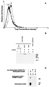
Figure 1
Figure 1
Binding of sCD26 to cell surface protein p300. (A) Specific binding of sCD26 to cell surface of K562 cells. Cells were stained with phycoerythrin-streptavidin alone (peak 1) or with biotinylated sCD26 and phycoerythrin-streptavidin (peak 2), followed by flow cytometry analysis. Binding of biotinylated sCD26 was inhibited by a 100-fold excess of nonbiotinylated sCD26 (peak 3). (B) Immunoprecipitation of cell surface CD26-binding protein p300. Surface-biotinylated K562 cells were incubated with sCD26 (lanes 2–5) or PBS alone (lane 1) followed by cross-linking with cleavable cross-linker DTSSP (20). sCD26 and its cross-linked complex were immunoprecipitated with three different anti-CD26 antibodies (lanes 1–4) or isotype-matched control antibody (TQ1; anti-l-selectin, lane 5). Immunoprecipitated materials were reduced (to cut cleavable chemical cross-linker) and then analyzed by SDS/PAGE and detected with streptavidin (20). (C) The binding of labeled sCD26 to p300 was inhibited by an excess amount of nonlabeled sCD26. K562 cells were incubated with biotinylated sCD26 (lanes 2 and 4) or PBS alone (lanes 1 and 3) in the presence (lanes 3 and 4) or absence (lanes 1 and 2) of a 100-fold excess of nonbiotinylated sCD26. After cross-linking with noncleavable cross-linker BS3, sCD26 and its cross-linked complex were immunoprecipitated by anti-CD26 antibody (1F7)-conjugated beads. Immunoprecipitated materials were electrophoresed and detected with streptavidin (20).
Figure 2
Figure 2
CD26-binding protein p300 is M6P/IGFIIR. (A and B) CD26-binding protein p300 was immunoprecipitated from surface-biotinylated K562 cells as described in Fig. 1B. After SDS/PAGE under reducing conditions and transfer to a membrane, p300 was detected with streptavidin (A). After stripping, the membrane was probed with rabbit anti-M6P/IGFIIR polyclonal antibody (B).
Figure 3
Figure 3
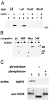
Binding of sCD26 and M6P/IGFIIR is mediated by M6P residues in the carbohydrate moiety of sCD26. (A and B) Binding of sCD26 and M6P/IGFIIR was inhibited by M6P. Surface-biotinylated K562 cells were incubated with sCD26 as in Fig. 1B in the presence of varying concentrations of M6P (A) or 100 μM M6P, mannose (Man), or G6P (B). M6P/IGFIIR was coprecipitated with sCD26 by using anti-CD26 antibody. (C) Requirement of glycosylation and phosphorylation of sCD26 for M6P/IGFIIR binding. sCD26 was incubated with buffer alone (lane 1), N- and O-glycosidases (lane 2), or alkaline phosphatase (lane 3) followed by SDS/PAGE and transfer to a membrane. Binding of enzyme-treated sCD26 and M6P/IGFIIR was analyzed by far Western blotting by using soluble M6P/IGFIIR as probe (upper blot). After stripping the membrane, sCD26 also was detected with Western blotting with anti-CD26 antibody (5F8) (lower blot).
Figure 4
Figure 4
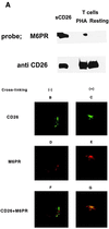
(A) Phytohemagglutinin activation of T cells increased the level of mannose 6 phosphorylation of intracellular CD26. CD26 was immunoprecipitated from the whole-cell lysate of resting or phytohemagglutinin-activated peripheral T cells. Mannose 6 phosphorylation of CD26 was analyzed by far Western blotting with M6P/IGFIIR as probe. Equal loading of CD26 was confirmed by Western blotting probed with anti-CD26 antibody (1F7). (BG) Immunocytochemical analysis of subcellular localization of CD26 and M6P/IGFIIR. Peripheral blood lymphocytes in resting state (B, D, and F) or stimulated by cross-linking of CD26 with anti-CD26 (C, E, and G) were stained with Oregon green 488 (green)-labeled anti-CD26 antibody (2F9) (B and C) and rabbit anti-M6P/IGFIIR antibody followed by rhodamine (red)-labeled anti-rabbit Ig antibody (16) (D and E). The stained cells were analyzed by confocal microscopy. Areas of coincidence of red and green fluorescence (giving yellow fluorescence) indicate overlapping distribution of CD26 and M6P/IGFIIR (F and H).
Figure 5
Figure 5
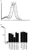
(A) The inhibitory effect of M6P on the modulation of CD26 by anti-CD26 antibody. Purified peripheral blood T cells were stained with phycoerythrin-conjugated anti-CD26 antibody (Ta1) before (line 1) or after (lines 2 and 3) cross-linking with anti-CD26 antibody (1F7) in the presence (line 3) or absence (line 2) of 2 mM M6P and analyzed by flow cytometry. Comparable results were obtained in five independent experiments. (B) The effect of M6P on T cell proliferation induced by CD3 and CD26 costimulation. Purified peripheral T cells were stimulated with anti-CD3 and anti-CD26 antibodies or anti-CD3 antibody with PMA in the presence or absence of M6P. In control experiments, G6P or mannose 1-phosphate was substituted for M6P. Proliferation was assessed by incorporation of [3H]thymidine into cells.

"V体育官网" References

    1. Morimoto C, Torimoto Y, Levinson G, Rudd C E, Schrieber M, Dang N H, Letvin N L, Schlossman S F. J Immunol. 1989;143:3430–3439. - PubMed
    1. Morimoto C, Schlossman S F. Immunol Rev. 1998;161:55–70. - PubMed
    1. Hegen M, Niedobitek G, Klein C E, Stein H, Fleischer B. J Immunol. 1990;144:2908–2914. - PubMed
    1. Torimoto Y, Dang N H, Tanaka T, Prado C, Schlossman S F, Morimoto C. Mol Immunol. 1992;29:183–192. - PubMed
    1. Oravecz T, Pall M, Roderiquez G, Gorrell M D, Ditto M, Nguyen N Y, Boykins R, Unsworth E, Norcross M A. J Exp Med. 1997;186:1865–1872. - PMC - PubMed

Publication types

MeSH terms