Skip to main page content
U.S. flag

An official website of the United States government

Dot gov

The . gov means it’s official VSports app下载. Federal government websites often end in . gov or . mil. Before sharing sensitive information, make sure you’re on a federal government site. .

Https

The site is secure V体育官网. The https:// ensures that you are connecting to the official website and that any information you provide is encrypted and transmitted securely. .

. 2000 Jul;20(14):5227-34.
doi: 10.1128/MCB.20.14.5227-5234.2000.

"V体育官网入口" c-Jun NH(2)-terminal kinase inhibits targeting of the protein phosphatase calcineurin to NFATc1

Affiliations

V体育平台登录 - c-Jun NH(2)-terminal kinase inhibits targeting of the protein phosphatase calcineurin to NFATc1

"V体育官网入口" C W Chow et al. Mol Cell Biol. 2000 Jul.

Abstract

The protein phosphatase calcineurin is a critical mediator of calcium signals during T-cell activation. One substrate of calcineurin is the transcription factor NFATc1, which is retained in the cytoplasm of quiescent cells. NFATc1 activation requires the translocation of the transcription factor into the nucleus, a process that is mediated by calcineurin. This interaction with calcineurin requires a targeting domain (PxIxIT motif) located in the NH(2)-terminal region of NFATc1 VSports手机版. Here we demonstrate that the calcineurin targeting domain of NFATc1 is phosphorylated and inactivated by the c-Jun NH(2)-terminal kinase (JNK). This disruption of calcineurin targeting inhibits the nuclear accumulation and transcription activity of NFATc1 and accounts for the observation that Jnk1(-/-) T cells exhibit greatly increased NFATc1-dependent nuclear responses. .


"VSports app下载" Figures

FIG. 1
FIG. 1
JNK1 inhibits NFAT activity in T cells. (A) Activation of JNK1 inhibits NFATc1 nuclear localization. Jurkat T cells were transfected without (empty expression vector; Control) and with MKK7 plus JNK1 (JNK). After 16 h, the cells were stimulated without (−) and with (+) ionomycin (I; 2 μM) plus PMA (P; 100 nM) for 6 h. Nuclear extracts were isolated (14), and NFATc1 was examined by protein immunoblot analysis using monoclonal antibody 7A6 (Affinity Bioreagents). Similar data were obtained in three independent experiments. (B) Activated JNK inhibits NFAT transcription activity. Jurkat T cells were transfected with an IL-4 promoter reporter gene (firefly luciferase) plasmid together without (Control) and with MKK7 plus JNK1 (JNK) or dnNFAT. Transfection efficiency was monitored by measurement of Renilla luciferase activity. The cells were stimulated without (□) and with (■) ionomycin (2 μM) plus PMA (100 nM) for 6 h. The data are presented as fold activation by in treated compared to untreated cells (mean ± standard deviation; n = 4).
FIG. 2
FIG. 2
Interaction of JNK1 with NFATc1-α. Lysates were prepared from COS cells transfected with HA-JNK1, and the binding of HA-JNK1 to immobilized recombinant NFATc1-α was examined. The effect NFATc1-α deletions is shown. The location of the JNK binding domain (JBD) and PxIxIT motif on NFATc1-α are illustrated schematically. Potential JNK phosphorylation sites (SP) are indicated.
FIG. 3
FIG. 3
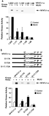
Phosphorylation of NFATc1-α by JNK1 in vitro. (A) JNK1 phosphorylates NFATc1-α. Immunocomplex kinase assays were performed using JNK1 activated without (Control; −) or with (+) MLK3 using recombinant NFATc1-α as the substrate. Phosphorylated NFATc1-α was detected by autoradiography and quantitated by PhosphorImager (Molecular Dynamics) analysis. (B) JNK phosphorylates NFATc1-α on Ser117 and Ser172. Wild-type and mutant NFATc1-α proteins (residues 1 to 202) were phosphorylated by JNK1 in vitro. The effects of JNK1 activation by MLK3 and the replacement of Ser117 and Ser172 with Ala were examined.
FIG. 4
FIG. 4
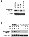
Phosphorylation of NFATc1-α in vivo. (A) NFATc1-α was coexpressed with MKK7 plus JNK1 in COS cells. The effect of replacement of Ser117 and Ser172 with Ala residues was investigated. Cell lysates were examined by protein immunoblot analysis by sequentially probing with anti-phospho-NFATc1-α and anti-NFATc1. A nonspecific band was detected by the phospho-NFATc1-α antibody immediately above the NFATc1-α band. (B) NFATc1-α was coexpressed without (−) and with (+) the JNK inhibitor JIP-1 in COS cells. The cells were treated without (−) and with (+) UV-C radiation (80 J/m2) and harvested after 30 min. The effect of replacement of the JNK phosphorylation sites (Ser117 and Ser172) with Ala was examined. The NFATc1-α proteins were detected by protein immunoblot analysis by sequentially probing with anti-phospho-NFATc1-α and anti-NFATc1.
FIG. 5
FIG. 5
JNK1 inhibits the nuclear accumulation of NFATc1-α. (A and B) Subcellular distribution of NFATc1-α, examined by immunofluorescence analysis in transfected BHK cells. The NFAT proteins (red; right) and nucleus (blue; left) were visualized (A). The effects of expression of a constitutively activated calcineurin (ΔCN) and MKK7 plus JNK1 (JNK) were examined. Arrowheads indicate the nuclei of cells expressing transfected proteins. The data were quantitated (B) following examination of 300 transfected cells and are presented as the percentage of cells with nuclear NFATc1-α (mean ± standard deviation [SD]; n = 3). (C) Effect of dnJNK on the subcellular distribution of NFATc1-α, examined by immunofluorescence analysis. The effects of expression of constitutively activated calcineurin (ΔCN) and MKK7 plus JNK1 (JNK) were examined. The percentage of cells with nuclear NFATc1-α is presented (mean ± SD; n = 3). (D) Effect of JIP-1 on the subcellular distribution of NFATc1-α, examined by immunofluorescence analysis. The percentage of cells with nuclear NFATc1-α is presented (mean ± SD; n = 3). The effects of increasing amounts of JIP-1 expression vector (0.3 and 0.7 μg) and the expression of constitutively activated calcineurin (ΔCN) and MKK7 plus JNK1 (JNK) were examined. (E) Mutational removal of Ser117 and Ser172 potentiates NFATc1-α transcription activity on the IL-4 promoter. Wild-type and [Ala117 Ala172] NFATc1-α were cotransfected with an IL-4 reporter plasmid in the absence (Untreated) and presence of MKK7 plus JNK1 (JNK). The cells were stimulated without (Untreated) and with PMA (P; 100 nM) plus ionomycin (I; 2 μM) for 16 h. The data are presented as fold activation compared to an untreated control (cells transfected without an NFATc1-α expression vector).
FIG. 6
FIG. 6
Differential regulation of NFATc1 isoforms by JNK. (A) The NH2-terminal regions of NFATc1 (18), NFATc1-α (25), and NFATc1-β (26, 33) are illustrated schematically. The PxIxIT motif (hatched box), the NFAT homology domain (NHD), and the alternative NFATc1 NH2 termini are indicated. Immobilized GST, GST-NFATc1 (residues 1 to 126), GST–NFATc1-α (residues 1 to 202), and GST–NFATc1-β (residues 1 to 189) was incubated with extracts prepared from COS cells transfected with HA-JNK1. The binding of HA-JNK1 was examined by immunoblot analysis. (B) Comparison of the phosphorylation of NFATc1 isoforms by JNK1 in vitro. Immunocomplex kinase assays were performed using Flag epitope-tagged JNK1 activated without (−) and with (+) MLK3, using recombinant NFATc1 isoforms as the substrate. (C and D) Subcellular distribution of NFATc1 isoforms, examined by immunofluorescence analysis in transfected BHK cells. The NFAT proteins (red; right) and nucleus (blue; left) were visualized (C). The effect of constitutively activated calcineurin (ΔCN) or MKK7 plus JNK1 (JNK) was examined. Arrowheads indicate the nuclei of cells expressing transfected proteins. The data were quantitated (D) following examination of 300 transfected cells and are presented as the percentage of cells with nuclear NFATc1 (mean ± standard deviation; n = 3).
FIG. 7
FIG. 7
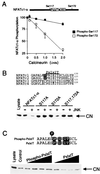
JNK1 inhibits the binding of calcineurin to NFATc1-α. (A) Comparison of calcineurin-stimulated dephosphorylion of Ser117 and Ser172. Recombinant NFATc1-α (residues 1 to 202) was phosphorylated by JNK1 in vitro and incubated (30 min at 30°C) in 50 mM HEPES (pH 7.4)–2 mM MnCl2–0.5 mM EDTA–15 mM 2-mercaptoethanol–0.1 mg of bovine serum albumin per ml together with calmodulin (250 U) and indicated amounts of calcineurin (Sigma). The phosphorylated NFATc1-α was detected after sodium dodecyl sulfate-polyacrylamide gel electrophoresis by autoradiography and was quantitated by PhosphorImager (Molecular Dynamics) analysis. Dephosphorylation at Ser117 and Ser172 was examined using [Ala172] and [Ala117] NFATc1-α, respectively. (B) Phosphorylation of NFATc1-α by JNK1 inhibits the binding of NFATc1-α to the phosphatase calcineurin. Recombinant NFATc1-α (residues 1 to 202) was phosphorylated by JNK1 in vitro. The NFATc1-α proteins were immobilized on GSH-Sepharose, incubated with cell extracts, and washed; bound calcineurin was detected by protein immunoblot analysis. The effect of replacement of the NFATc1-α phosphorylation sites (Ser117 and Ser172) with Ala was examined. (C) Phosphorylation of Ser117 inhibits calcineurin binding to NFATc1-α. Immobilized recombinant NFATc1-α (residues 1 to 202) was incubated with cell extracts, and the binding of calcineurin was examined by protein immunoblot analysis. Competition analysis was performed by investigating the effects of various concentrations (0, 0.7, 1.4, 7, and 14 μM) of a synthetic peptide corresponding to the PxIxIT motif which mediates the targeting of calcineurin to NFATc1-α. The effect of the phosphorylation of the synthetic peptide on Ser117 was examined.
FIG. 8
FIG. 8
NFATc1-α binding to CRM1 is regulated by phosphorylation. (A) Activated calcineurin increases the binding of NFATc1-α to CRM1. NFATc1-α was expressed without (−) or with (+) activated calcineurin (ΔCN) in COS cells. Recombinant CRM1 was immobilized on GSH-Sepharose, incubated with the COS cell extracts, and washed; bound NFATc1-α was detected by protein immunoblot analysis. (B) JNK phosphorylation inhibits the binding of NFATc1-α to CRM1. The effect of replacement of the JNK phosphorylation sites (Ser117 and Ser172) on the binding of NFATc1-α to CRM1 was examined. The effect of JNK inhibition was investigated by overexpression of the scaffold protein JIP-1.

References

    1. Aramburu J, Garcia-Cozar F, Raghavan A, Okamura H, Rao A, Hogan P G. Selective inhibition of NFAT activation by a peptide spanning the calcineurin targeting site of NFAT. Mol Cell. 1998;1:627–637. - PubMed (V体育2025版)
    1. Aramburu J, Yaffe M B, Lopez-Rodriguez C, Cantley L C, Hogan P G, Rao A. Affinity-driven peptide selection of an NFAT inhibitor more selective than cyclosporin A. Science. 1999;285:2129–2133. - PubMed
    1. Avots A, Buttmann M, Chuvpilo S, Escher C, Smola U, Bannister A J, Rapp U R, Kouzarides T, Serfling E. CBP/p300 integrates Raf/Rac-signaling pathways in the transcriptional induction of NF-ATc during T cell activation. Immunity. 1999;10:515–524. - "VSports最新版本" PubMed
    1. Beals C R, Clipstone N A, Ho S N, Crabtree G R. Nuclear localization of NF-ATc by a calcineurin-dependent, cyclosporin-sensitive intramolecular interaction. Genes Dev. 1997;11:824–834. - PubMed
    1. Beals C R, Sheridan C M, Turck C W, Gardner P, Crabtree G R. Nuclear export of NF-ATc enhanced by glycogen synthase kinase-3. Science. 1997;275:1930–1934. - "VSports在线直播" PubMed

Publication types

"VSports最新版本" MeSH terms

Substances

"V体育平台登录" LinkOut - more resources