Skip to main page content (V体育2025版)
U.S. flag

An official website of the United States government

Dot gov

The . gov means it’s official. Federal government websites often end in . gov or . mil. Before sharing sensitive information, make sure you’re on a federal government site VSports app下载. .

Https

The site is secure. The https:// ensures that you are connecting to the official website and that any information you provide is encrypted and transmitted securely V体育官网. .

. 2000 Jun;20(12):4309-19.
doi: 10.1128/MCB.20.12.4309-4319.2000.

The c-Myc transactivation domain is a direct modulator of apoptotic versus proliferative signals

Affiliations

The c-Myc transactivation domain is a direct modulator of apoptotic versus proliferative signals

D W Chang et al. Mol Cell Biol. 2000 Jun.

Abstract

We have assayed the oncogenic, proliferative, and apoptotic activities of the frequent mutations that occur in the c-myc gene in Burkitt's lymphomas. Some alleles have a modest (50 to 60%) increase in transforming activity; however, the most frequent Burkitt's lymphoma allele (T58I) had an unexpected substantial decrease in transforming activity (85%). All alleles restored the proliferation function of c-Myc in cells that grow slowly due to a c-myc knockout. There was discordance for some alleles between apoptotic and oncogenic activities, but only the T58A allele had elevated transforming activity with a concomitant reduced apoptotic potential. We discovered a novel missense mutation, MycS71F, that had a very low apoptotic activity compared to wild-type Myc, yet this mutation has never been found in lymphomas, suggesting that there is no strong selection for antiapoptotic c-Myc alleles. MycS71F also induced very low levels of cytochrome c release from mitochondria, suggesting a mechanism of action for this mutation. Phosphopeptide mapping provided a biochemical basis for the dramatically different biological activities of the transformation-defective T58I and transformation-enhanced T58A c-Myc alleles. Furthermore, the antiapoptotic survival factor insulin-like growth factor 1 was found to suppress phosphorylation of T58, suggesting that the c-Myc transactivation domain is a direct target of survival signals. VSports手机版.


"V体育ios版" Figures
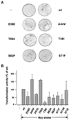
FIG. 1
FIG. 1
Transformation activity of wt and mutant c-myc genes in the rat embryo fibroblast focus assay. (A) Representative pictures of the rat embryo fibroblast monolayers with H-rasG12V only (−) or H-rasG12V with wt or mutant c-myc. (B) Relative transformation activities of various mutant c-myc alleles as a percentage of wt c-myc activity. Data shown are averages of two to three independent experiments. Differences in transforming activity are statistically significant based on P values of <0.05 using the Student t test. Protein expression was equal for all alleles after transient transfection into rat embryo fibroblasts (data not shown).
FIG. 2
FIG. 2
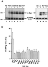
Reconstitution of c-Myc protein expression and cell proliferation in c-myc null cells. (A) Wild-type or mutant c-myc reconstituted cell lines have similar levels of c-Myc protein, but the level is slightly higher than that in the c-myc diploid TGR cells. c-Myc proteins were immunoprecipitated from equal amounts of cycling cell lysate and detected by Western blotting. Ig, immunoglobulin. Sizes are given in kilodaltons. (B) Average doubling time of fibroblast cell lines expressing either no c-Myc (HO), endogenous diploid levels of c-Myc (TGR), or wt or mutant c-Myc in reconstituted cell lines. Bars represent doubling times ± standard error for three to five experiments, and two to four clones were assayed for each reconstituted allele. The difference in doubling time between wt Myc and T58I or S62A reconstituted lines is statistically significant based on P values of <0.02 using the Student t test.
FIG. 3
FIG. 3
Apoptotic versus transforming activity with differing c-Myc mutations. (A) Representative flow cytometry profiles of HO, TGR, and various reconstituted cell lines following serum starvation (0.1%) for 24 h. DNA content was measured by fluorescence with propidium iodide staining. M1, M2, M3, and M4 represent gating for G1, S, G2, and sub-G1 populations, respectively. (B) Histogram of average percent apoptosis of the various cell lines in low serum as calculated by FACS gating for the sub-G1 population (M4). Data represent averages of three to five independent determinations. The oncogenic activity of each mutant c-Myc protein from Fig. 1B is shown for comparison.
FIG. 3
FIG. 3
Apoptotic versus transforming activity with differing c-Myc mutations. (A) Representative flow cytometry profiles of HO, TGR, and various reconstituted cell lines following serum starvation (0.1%) for 24 h. DNA content was measured by fluorescence with propidium iodide staining. M1, M2, M3, and M4 represent gating for G1, S, G2, and sub-G1 populations, respectively. (B) Histogram of average percent apoptosis of the various cell lines in low serum as calculated by FACS gating for the sub-G1 population (M4). Data represent averages of three to five independent determinations. The oncogenic activity of each mutant c-Myc protein from Fig. 1B is shown for comparison.
FIG. 4
FIG. 4
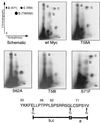
Thermolytic phosphopeptide mapping analysis of Myc null cells stably expressing wt c-Myc or c-Myc proteins with phosphorylation point mutations, as indicated. Cells were labeled for 4 h with [32P]orthophosphate. Myc proteins were immunoprecipitated and processed for 2D phosphopeptide mapping as described in Materials and Methods. The schematic representation of the 2D thermolytic peptide map (top left) indicates major sites of phosphorylation at S71 (peptide a), T58/S62 (peptide b), and S62 (peptide c) following thermolytic digestion. The diagram at the bottom represents predicted thermolytic peptides a, b, and c. Arrows indicate thermolysin (Th) digestion sites.
FIG. 5
FIG. 5
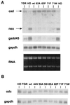
c-Myc proteins induce equivalent activation and repression of target genes. (A) Northern blot of the parental and reconstituted cell line RNAs sequentially probed for cad, neo, gadd45, and gapdh RNAs. The ethidium bromide-stained gel is shown in the lower panel. (B) Northern blot of odc and gapdh expression as above.
FIG. 6
FIG. 6
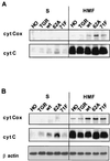
c-Myc-induced apoptotic activity correlates with cytochrome c release from mitochondria. (A) Cycling c-myc null cells (HO), c-myc diploid cells (TGR), and null cells reconstituted with wt c-Myc, MycS62A, and MycS71F were lysed in an isotonic sucrose buffer by Dounce homogenization and fractionated by centrifugation. Equivalent amounts of protein from the resultant supernatant and pellet (HMF) were separated by SDS-PAGE, blotted onto a nitrocellulose membrane, and probed with anti-cytochrome c (cyt C) or anti-cytochrome c oxidoreductase (cyt Cox). The latter protein serves as a control for mitochondrial integrity. (B) Cytochrome c release from the above cells in 0.1% serum following the same lysis protocol. After transfer, the blot was probed with antibodies for cytochrome c and cytochrome c oxidoreductase. To verify equal protein loading, the same blot was reprobed with anti-β-actin (bottom panel).
FIG. 7
FIG. 7
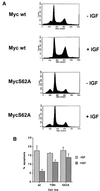
IGF-1 rescue of c-Myc-induced apoptosis is mutation specific. (A) Flow cytometry of wt Myc and MycS62A reconstituted cells cultured for 18 h in 0.1% serum in the presence or absence of IGF-1 (200 ng/ml). Both floating and attached cells were harvested, fixed, and stained with propidium iodide before determination of the DNA content by flow cytometry. (B) Apoptotic activity of different c-Myc mutants after gating for the sub-G1 population in serum-starved cells with or without IGF-1. Data represent the averages of three independent experiments ± standard error.
FIG. 8
FIG. 8
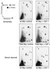
IGF-1 suppresses the phosphorylation of c-Myc T58, as determined by thermolytic phosphopeptide mapping analysis of Myc null cells stably expressing either wt c-Myc or MycT58A. Cells were untreated or treated with IGF-1 (200 ng/ml) for 20 h prior to labeling for 4 h with [32P]orthophosphate. The top and middle panels were derived from log-phase cells in 10% serum, whereas the bottom panels were derived from cells starved in 0.1% serum for 20 h before labeling. Myc proteins were immunoprecipitated and processed for 2D phosphopeptide mapping as described in Materials and Methods. The schematic representation of the 2D thermolytic peptide map (top left) indicates major sites of phosphorylation at S71 (peptide a), T58/S62 (peptide b), and S62 (peptide c) following thermolytic digestion. Peptide c resolved into two closely spaced spots in the serum-starved IGF-1 treated cells, but this was not found in other experiments.

References

    1. Albert T, Urlbauer B, Kohlhuber F, Hammersen B, Eick D. Ongoing mutations in the N-terminal domain of c-Myc affect transactivation in Burkitt's lymphoma cell lines. Oncogene. 1994;9:759–763. - PubMed
    1. Alverez E, Northwood I, Gonzalez F, Latour D, Seth A, Abate C, Curran T, Davis R. Pro-Leu-Ser/Thr-Pro is a consensus primary sequence for substrate protein phosphorylation. Characterization of the phosphorylation of c-myc and c-jun proteins by an epidermal growth factor receptor threonine 669 protein kinase. J Biol Chem. 1991;266:15277–15285. - PubMed
    1. Askew D S, Ashmun R A, Simmons B C, Cleveland J L. Constitutive c-myc expression in an IL-3-dependent myeloid cell line suppresses cell cycle arrest and accelerates apoptosis. Oncogene. 1991;6:1915–1922. - PubMed
    1. Beijersbergen R L, Hijmans E M, Zhu L, Bernards R. Interaction of c-Myc with the pRb-related protein p107 results in inhibition of c-Myc-mediated transactivation. EMBO J. 1994;13:4080–4086. - "V体育安卓版" PMC - PubMed
    1. Bhatia K, Huppi K, Spangler G, Siwarski D, Iyer R, Magrath I. Point mutations in the c-Myc transactivation domain are common in Burkitt's lymphoma and mouse plasmacytomas. Nat Genet. 1993;5:56–61. - PubMed

Publication types