Skip to main page content
U.S. flag

An official website of the United States government

Dot gov

The . gov means it’s official. Federal government websites often end in . gov or . mil. Before sharing sensitive information, make sure you’re on a federal government site VSports app下载. .

Https

The site is secure V体育官网. The https:// ensures that you are connecting to the official website and that any information you provide is encrypted and transmitted securely. .

. 2000 May 9;97(10):5101-6.
doi: 10.1073/pnas.97.10.5101.

Glutathione synthesis is essential for mouse development but not for cell growth in culture

Affiliations

Glutathione synthesis is essential for mouse development but not for cell growth in culture

V体育安卓版 - Z Z Shi et al. Proc Natl Acad Sci U S A. .

Abstract

Glutathione (GSH) is a major source of reducing equivalents in mammalian cells. To examine the role of GSH synthesis in development and cell growth, we generated mice deficient in GSH by a targeted disruption of the heavy subunit of gamma-glutamylcysteine synthetase (gammaGCS-HS(tm1)), an essential enzyme in GSH synthesis VSports手机版. Embryos homozygous for gammaGCS-HS(tm1) fail to gastrulate, do not form mesoderm, develop distal apoptosis, and die before day 8. 5. Lethality results from apoptotic cell death rather than reduced cell proliferation. We also isolated cell lines from homozygous mutant blastocysts in medium containing GSH. These cells also grow indefinitely in GSH-free medium supplemented with N-acetylcysteine and have undetectable levels of GSH; further, they show no changes in mitochondrial morphology as judged by electron microscopy. These data demonstrate that GSH is required for mammalian development but dispensable in cell culture and that the functions of GSH, not GSH itself, are essential for cell growth. .


Figures

Figure 1
Figure 1
Structure of the γ-GCS-HS gene and targeted disruption of γ-GCS-HS. (A) Structure of the mouse γGCS-HS locus. Exons (solid boxes) and introns (solid line) are drawn to scale. The positions of the nucleotides at the 3′ limits of exons 1–15 are as follows: 150, 299, 482, 596, 655, 789, 864, 981, 1120, 233, 1326, 1431, 1504, 1617, and 1738 (see ref. for cDNA sequence). (B) Targeting scheme for γGCS-HS disruption. The genomic structure of the 5′ portion of the gene is presented at the top. The targeting vector (middle) was designed such that the PGK-hprt cassette replaced exon 1. An MC1-tk cassette was used as a negative selectable marker. The predicted mutant allele is shown at the bottom. Two external probes used to determine targeting events are indicated as slashed boxes. They both were expected to hybridize with shortened “mutant” bands from the mutant allele. Restriction sites: B, BamHI; E, EcoRI; H, HindIII. (C) Southern blot analysis of genomic DNA isolated from mouse tails derived from heterozygote matings. Only the analysis using the 5′ probe is shown. (D) PCR genotype analysis of mouse embryos. Each DNA sample was subjected to two separate PCRs using the primer pairs designed for either the wild-type (WT) or the mutant band. Two primer pairs are indicated with arrows in B.
Figure 2
Figure 2
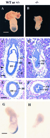
Developmental abnormalities in E7.5 γGCS-HS mutant embryos. (A and B) Whole-mount preparations of E7.5 normal and mutant embryos. The mutant embryos (B) are smaller than normal (A) and show lack of organization. (CF) Histological comparison of normal (C and E) and mutant (D and F) embryos. The arrowheads in the sagittal sections (C and D) indicate the approximate position of the transverse sections (E and F). Note lack of mesoderm and ectoplacental, exocoelomic, and amniotic cavities in the mutant embryos. d, Decidua; ee, embryonic ectoderm; m, mesoderm; ve, visceral endoderm. (G and H) Whole-mount in situ hybridization analysis with a mesoderm marker, Brachyury (T). A normal expression pattern of T gene is shown in the wild-type embryo (G). No signal was detected in a mutant littermate (H). [Bar: 300 μm (A, B, G, and H); 100 μm (CF).]
Figure 3
Figure 3
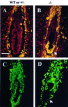
In vivo apoptosis and proliferation in E6.5 γGCS-HS mutant embryos. (A and B) Sagittal sections from two embryos [wild-type (WT) or heterozygous (+/−) and homozygous mutant (−/−)] were assayed by the TUNEL reaction (ApopTaq fluorescein kit, Oncor/Intergen). Fluorescein-labeled nuclei (orange) indicate apoptotic cells. Unlabeled nuclei appear red as a result of counterstaining with propidium iodide. The normal embryo (A) shows few apoptotic nuclei, whereas the mutant embryo (B) shows severe distal apoptosis (arrow). (C and D) Sagittal sections from wild-type or +/− and −/− littermate embryos were analyzed by BrdUrd incorporation. Positive nuclei are visualized by green fluorescence. The mutant embryo (D) shows total absence of BrdUrd incorporation at its distal end (arrow), but the incorporation in other regions is comparable to the embryo (C). Genotypes of embryos were determined in adjacent sections by in situ hybridization with a γGCS-HS exon 1 probe (not shown). (Bar: 100 μM.)
Figure 4
Figure 4
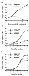
Growth of γGCS-HS-deficient cells. (A) Dependence of GCS-1 cells on exogenous GSH for growth. Cells were seeded in 6-well plates in duplicate with M15 medium containing 2.5 mM GSH. On day 0 they were refed with M15 medium with or without GSH. Trypan blue-excluding cells were counted at 24-h intervals. (B) Support of GCS-1 cell growth by NAC. On day 0, cells were seeded in M15 medium containing 2.5 mM GSH or 2 or 5 mM of NAC in place of GSH. (C) Growth of GCS-1nac subline cells in the presence of NAC. Cells were seeded in M15 medium containing the additions shown. In all experiments, medium was changed daily.
Figure 5
Figure 5
Determination of cellular GSH by HPLC/ECD. (A) Chromatographic profile of acid soluble extract of BDC-1 (wild-type) cells. (B) Portions of chromatograms expanded between 7.5 and 9.5 min to show the presence and absence of GSH in γGCS-deficient cells: 1, GCS-1 cells grown in the presence of 2.5 mM GSH; 2, GCS-1 cells after withdrawal of GSH from the medium for 24 h; 3, GCS-1nac cells grown in the presence of 2 mM NAC. Note absence of GSH peak in 2 or 3. The small peak to the right of the GSH peak is an unidentified compound found in all cells. Note the difference in the μA scales for A and B.

References

    1. Kosower N S. Int Rev Cytol. 1978;54:109–160. - PubMed (VSports最新版本)
    1. Meister A. J Biol Chem. 1988;263:17205–17208. - PubMed
    1. Holmgren A. In: Glutathione: Metabolism and Physiological Function. Viña J, editor. Boca Raton, FL: CRC; 1990. pp. 145–154.
    1. Luthman M, Eriksson S, Holmgren A, Thelander L. Proc Natl Acad Sci USA. 1979;76:2158–2162. - PMC - PubMed
    1. Prinz W A, Aslund F, Holmgren A, Beckwith J. J Biol Chem. 1997;272:15661–15667. - PubMed

Publication types

MeSH terms