<kbd date-time="MzLmW"></kbd><del id="Q9F64z"></del> "V体育官网入口" Skip to main page content
U.S. flag

An official website of the United States government

Dot gov

The . gov means it’s official. Federal government websites often end in . gov or . mil. Before sharing sensitive information, make sure you’re on a federal government site. VSports app下载.

Https

The site is secure V体育官网. The https:// ensures that you are connecting to the official website and that any information you provide is encrypted and transmitted securely. .

. 2000 Feb 1;97(3):1263-8.
doi: 10.1073/pnas.97.3.1263.

"V体育官网" Tyrosine phosphorylation of the Helicobacter pylori CagA antigen after cag-driven host cell translocation

Affiliations

Tyrosine phosphorylation of the Helicobacter pylori CagA antigen after cag-driven host cell translocation

M Stein et al. Proc Natl Acad Sci U S A. .

Abstract (VSports手机版)

Helicobacter pylori strains associated with severe tissue damage and inflammation possess a unique genetic locus, cag, containing 31 genes originating from a distant event of horizontal transfer and retained as a pathogenicity island VSports手机版. The cag system is an Helicobacter-specific type IV secretion engine involved in cellular responses like induction of pedestals, secretion of IL-8, and phosphorylation of proteic targets. It has previously been reported that cocultivation of epithelial cells with Helicobacter pylori triggers signal transduction and tyrosine phosphorylation of a 145-kDa putative host cell protein. Herein, we demonstrate that this protein is not derived from the host but rather is the bacterial immunodominant antigen CagA, a virulence factor commonly expressed in peptic ulcer disease and thought to be an orphan of a specific biological function. Thus, CagA is delivered into the epithelial cells by the cag type IV secretion system where it is phosphorylated on tyrosine residues by an as yet unidentified host cell kinase and wired to eukaryotic signal transduction pathways and cytoskeletal plasticity. .


Figures

Figure 1
Figure 1
Radioactive profile of tyrosine-phosphorylated proteins after infection of AGS cells with 35S-labeled Hp. AGS cells were infected with the 35S-labeled Hp strain 87A300. Tyrosine-phosphorylated proteins were immunoprecipitated from RIPA-buffer-soluble fractions by using monoclonal anti-phosphotyrosine antibody (anti-PY; PY20, Transduction Laboratories, Lexington, KY). Immunoprecipitates (imp) and proteins of the RIPA-soluble cell fraction after the immunoprecipitation with anti-PY (post-imp) were separated by SDS/6% PAGE and transferred onto nitrocellulose membranes in duplicate. One membrane was exposed to Kodak x-ray film (A); the second nitrocellulose membrane was probed with anti-phosphotyrosine antibody (anti-PY). Blots were developed with peroxidase-coupled secondary antibodies [GIBCO/BRL]. The arrow in A indicates the 35S-labeled protein immunoprecipitated with anti-PY and comigrating with PTYR (shown in B).
Figure 2
Figure 2
Anti-CagA antibodies recognize PTYR. AGS cells were infected with the Hp strain 87A300. Lysis (lys), fractionation, and immunoprecipitation (imp) of infected (AGS+Hp) and uninfected (AGS) tissue culture cells were performed as described for Fig. 1. The fractions were analyzed with different antibodies: polyclonal human anti-Hp (A), polyclonal anti-CagA serum and preserum (pre; B), and monoclonal anti-CagA antibody (LDS 56; C). All three antibodies recognized the same bands including immunoprecipitated PTYR. (post imp, postimmunoprecipitation.) (D) CagA immunoprecipitated from infected cells with polyclonal anti-CagA serum (imp α-CagA) was recognized with anti-PY.
Figure 3
Figure 3
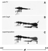
Two-dimensional gel electrophoresis showing the similar migration pattern of PTYR and CagA. Infected AGS cells were denatured in electrophoresis buffer. After analysis by two-dimensional gel electrophoresis (16), the proteins were transferred onto poly(vinylidene difluoride) membrane. The membrane was probed with anti-PY (A) and then stripped and reprobed with polyclonal anti-CagA antibody (B). (C) A superimposition of A and B. Arrows in B point to the unphosphorylated form of CagA, and the major proteic spot at higher pI is indicated by a bold arrow. The CagA spots with slightly lower molecular masses are indicated with regular arrows. Arrowheads in C indicate a degradation product.
Figure 4
Figure 4
The molecular mass of PTYR changes according to the molecular mass of the CagA antigen. Wild-type (wt) strains G27 and 342 and their isogenic cagA knockout mutants were incubated with AGS cells (12). Proteins were analyzed by SDS/6% PAGE and probed with anti-PY or with monoclonal anti-CagA antibody (LDS 56) by Western blotting. The mobility of CagA was estimated by SDS/PAGE and was also inferred by DNA sequence analysis of the 102-bp repeat region. The presence of two repeats in strain G27 and one repeat in strain 342 allowed precise calculation of the size of CagA and explains the kDa difference.
Figure 5
Figure 5
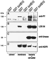
PTYR fractionation. AGS cells were infected with the Hp wild-type strain G27 or its isogenic mutant Δ527 as described (12). Host cell cytosol and membrane proteins were separated from the cellular fraction containing the bacteria, unlysed cells, and the cytoskeleton proteins. The fractions were analyzed by SDS/8% PAGE and transferred onto four separate nitrocellulose membranes. Each membrane was probed with a different antibody [anti-PY (A), LDS 56 (B), anti-urease (rabbit polyclonal antiserum against recombinant UreA; C), or anti-HGFR (C28, Santa Cruz Biotechnology; D)]. Estimated mobility is provided for the phosphorylated (A; 138 kDa) and unphosphorylated CagA (B; 136 kDa), urease A (C; 58 kDa), and hepatocyte growth factor receptor (HGFR) major subunit (D; 145 kDa).
Figure 6
Figure 6
Model for Helicobacter-mediated signaling events. (A) CagA (schematically drawn as a circle with a tyrosine residue, Y) is translocated via the type IV secretion system, represented as channels. (B) Black triangles represent cellular kinases that phosphorylate CagA on the tyrosine residue (the phosphate group is indicated by *P). CagA triggers signals that induce pedestal formation. In C, the pedestal elongates.

References

    1. Covacci A, Telford J L, Del Giudice G, Parsonnet J, Rappuoli R. Science. 1999;284:1328–1333. - PubMed
    1. Papini E, Satin B, Bucci C, de Bernard M, Telford J L, Manetti R, Rappuoli R, Zerial M, Montecucco C. EMBO J. 1997;16:15–24. - PMC (VSports手机版) - PubMed
    1. Papini E, Gottardi E, Satin B, de Bernard M, Massari P, Telford J, Rappuoli R, Sato S B, Montecucco C. J Med Microbiol. 1996;45:84–89. - PubMed
    1. Alm R A, Ling L S, Moir D T, King B L, Brown E D, Doig P C, Smith D R, Noonan B, Guild B C, deJonge B L, et al. Nature (London) 1999;397:176–180. - PubMed (VSports注册入口)
    1. Tomb J F, White O, Kerlavage A R, Clayton R A, Sutton G G, Fleischmann R D, Ketchum K A, Klenk H P, Gill S, Dougherty B A, et al. Nature (London) 1997;388:539–547. - PubMed (V体育安卓版)

Publication types

MeSH terms

VSports app下载 - LinkOut - more resources