Skip to main page content
U.S. flag

An official website of the United States government

Dot gov

The . gov means it’s official. Federal government websites often end in . gov or . mil. Before sharing sensitive information, make sure you’re on a federal government site VSports app下载. .

Https

The site is secure. The https:// ensures that you are connecting to the official website and that any information you provide is encrypted and transmitted securely. V体育官网.

. 1999 Nov 9;96(23):13363-8.
doi: 10.1073/pnas.96.23.13363.

VSports手机版 - Interaction of the copper chaperone HAH1 with the Wilson disease protein is essential for copper homeostasis

Affiliations

Interaction of the copper chaperone HAH1 with the Wilson disease protein is essential for copper homeostasis

I Hamza et al. Proc Natl Acad Sci U S A. .

Abstract

The delivery of copper to specific sites within the cell is mediated by distinct intracellular carrier proteins termed copper chaperones. Previous studies in Saccharomyces cerevisiae suggested that the human copper chaperone HAH1 may play a role in copper trafficking to the secretory pathway of the cell. In this current study, HAH1 was detected in lysates from multiple human cell lines and tissues as a single-chain protein distributed throughout the cytoplasm and nucleus. Studies with a glutathione S-transferase-HAH1 fusion protein demonstrated direct protein-protein interaction between HAH1 and the Wilson disease protein, which required the cysteine copper ligands in the amino terminus of HAH1. Consistent with these in vitro observations, coimmunoprecipitation experiments revealed that HAH1 interacts with both the Wilson and Menkes proteins in vivo and that this interaction depends on available copper. When these studies were repeated utilizing three disease-associated mutations in the amino terminus of the Wilson protein, a marked diminution in HAH1 interaction was observed, suggesting that impaired copper delivery by HAH1 constitutes the molecular basis of Wilson disease in patients harboring these mutations VSports手机版. Taken together, these data provide a mechanism for the function of HAH1 as a copper chaperone in mammalian cells and demonstrate that this protein is essential for copper homeostasis. .


Figures
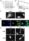
Figure 1
Figure 1
(A) Immunoblot analysis with HAH1 antibody. Protein lysates (100 μg) (lanes 1–4) or 100 ng of recombinant HAH1 expressed in E. coli (lane 5) was separated by SDS/PAGE on 10–20% Tricine gradient gels, transferred to nitrocellulose, and analyzed by chemiluminescence. (B) HeLa cells grown in the presence of BCS (●) or copper (○) were lysed and immunoprecipitated with HAH1 antisera. Following a 2-hr pulse with [35S]methionine and [35S]cysteine, cells were lysed and immunoprecipitated at indicated chase times followed by SDS/PAGE, autoradiography, and quantitation of bands. (Inset) HAH1 immunoprecipitated in the presence of BCS. (C) Immunofluorescent localization of HAH1 in HeLa cells incubated with HAH1 antisera (α-HAH1; confocal 100X) and Flag M2 antibody (α-Flag; 60X). (D) Immunofluorescence of HeLa cells stained for nucleus with DAPI (narrow-band UV), HAH1 antiserum (FITC), or overlay images of DAPI and HAH1. (E) Double immunofluorescence in transfected HeLa cells treated with BCS (−Cu) or copper (+Cu) followed by incubation with Menkes (α-Mnk) and Flag-M2 (α-Flag) antibodies. Arrows indicate transfected HeLa cells.
Figure 2
Figure 2
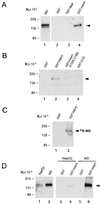
In vitro interaction of HAH1 and Wilson protein. (A) COS-7 cell lysates (150 μg) expressing Wilson protein (5 μg; lane 1) were incubated with 50 pmol of purified GST-fusion proteins (lanes 2–4), and eluates were subjected to SDS/PAGE, transferred to nitrocellulose, and examined by immunoblotting with Wilson antisera. (B) COS-7 cell lysates expressing the Wilson protein were incubated with the indicated GST constructs (lanes 1–4) and analyzed as indicated above. (C) Full length Wilson cDNA was translated in vitro in the presence of [35S]methionine and [35S]cysteine followed by incubation with the GST constructs as indicated. Interacting proteins were separated by SDS/PAGE and analyzed by autoradiography. (D) One milligram of HepG2 cell lysate (100 μg; lane 1) or 150 μg COS-7 cell lysate expressing the Wilson protein (5 μg; lane 2) was incubated with 3 nmol of purified GST fusion proteins (lanes 3–6) and analyzed by immunoblotting with Wilson antisera as before.
Figure 3
Figure 3
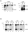
(A) HepG2 cells were immunoprecipitated with either HAH1, Wilson, CCS, or SOD1 antisera, and the immunoprecipitates were separated by SDS/PAGE transferred to nitrocellulose and examined by immunoblotting with HAH1 (α-HAH1) or Wilson (α-WD) antisera. (B) HeLa and HepG2 cells grown in the presence of 200 μM copper (lanes 1, 2 and 5, 6) or 50 μM BCS (lanes 3, 4 and 7, 8) were immunoprecipitated with HAH1 (lanes 1, 3, 5, 7), Menkes (lanes 2, 4) or Wilson (lanes 6, 8) antisera. Immunoprecipitates were analyzed with HAH1 antisera as indicated above. IgG heavy and light chains detected by anti-rabbit secondary antibody are indicated.
Figure 4
Figure 4
(A) Amino acid sequence alignment of HAH1 and the copper-binding domains of the Wilson (WD), Menkes (MK), and RAN-1 proteins. The conserved MXCXXC motif and glycine residue are outlined. The Wilson disease mutations (G85V, L492S and G591D) are highlighted, and the RAN-1Cu2 mutation (G173E) noted by asterisk. (B) Structural model (Insight II, Molecular Simulations) of the Menkes fourth copper-binding domain on the basis of the NMR structure (16) depicting the relative positions of the Wilson protein and RAN-1 mutations. C388 and C391 are the copper-binding cysteines. Hydrogen bonds (Å) between I381 and G417 in an antiparallel β-sheet of the Menkes protein are indicated and correspond to L492 in the Wilson protein and G173 in RAN-1, respectively. G404 is shown with van der Waals surface and corresponds to the G85 and G591 Wilson protein mutations.
Figure 5
Figure 5
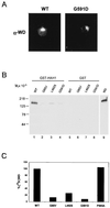
(A) Indirect immunofluorescence of Wilson protein in COS-7 cells transiently transfected with wild type (Left) or G591D mutant (Right). Cells were processed for immunofluorescence 72 hr posttransfection (X60). (B) COS-7 cells expressing either wild-type or mutant Wilson proteins were lysed, incubated with GST-HAH1, and interacting proteins were separated by SDS/PAGE and analyzed by immunoblotting with Wilson antisera. (C) Wild-type and mutant Wilson proteins were translated in vitro in the presence of [35S]methionine and [35S]cysteine, incubated with GST-HAH1, and interacting proteins analyzed by fluorography after SDS/PAGE and were quantitated by PhosphorImager.

V体育官网 - References

    1. Solomon E I, Lowery M D. Science. 1993;259:1575–1581. - PubMed (V体育官网)
    1. Harris Z L, Gitlin J D. Am J Clin Nutr. 1996;63:836S–841S. - PubMed
    1. Schaefer M, Gitlin J D. Am J Physiol. 1999;276:G311–G314. - PubMed
    1. Valentine J S, Gralla E B. Science. 1997;278:817–818. - V体育官网入口 - PubMed
    1. Lin S J, Culotta V C. Proc Natl Acad Sci USA. 1995;92:3784–3788. - PMC - PubMed

Publication types

VSports注册入口 - MeSH terms

Substances (VSports最新版本)

LinkOut - more resources (V体育平台登录)