"V体育2025版" Skip to main page content
U.S. flag

An official website of the United States government

Dot gov

The . gov means it’s official. Federal government websites often end in . gov or . mil. Before sharing sensitive information, make sure you’re on a federal government site. VSports app下载.

Https

The site is secure. The https:// ensures that you are connecting to the official website and that any information you provide is encrypted and transmitted securely V体育官网. .

. 1999 Nov 9;96(23):13318-23.
doi: 10.1073/pnas.96.23.13318.

Specific binding of proinsulin C-peptide to human cell membranes

Affiliations

Specific binding of proinsulin C-peptide to human cell membranes

R Rigler et al. Proc Natl Acad Sci U S A. .

Abstract

Recent reports have demonstrated beneficial effects of proinsulin C-peptide in the diabetic state, including improvements of kidney and nerve function. To examine the background to these effects, C-peptide binding to cell membranes has been studied by using fluorescence correlation spectroscopy. Measurements of ligand-membrane interactions at single-molecule detection sensitivity in 0. 2-fl confocal volume elements show specific binding of fluorescently labeled C-peptide to several human cell types. Full saturation of the C-peptide binding to the cell surface is obtained at low nanomolar concentrations. Scatchard analysis of binding to renal tubular cells indicates the existence of a high-affinity binding process with K(ass) > 3 VSports手机版. 3 x 10(9) M(-1). Addition of excess unlabeled C-peptide is accompanied by competitive displacement, yielding a dissociation rate constant of 4. 5 x 10(-4) s(-1). The C-terminal pentapeptide also displaces C-peptide bound to cell membranes, indicating that the binding occurs at this segment of the ligand. Nonnative D-C-peptide and a randomly scrambled C-peptide do not compete for binding with the labeled C-peptide, nor were crossreactions observed with insulin, insulin-like growth factor (IGF)-I, IGF-II, or proinsulin. Pretreatment of cells with pertussis toxin, known to modify receptor-coupled G proteins, abolishes the binding. It is concluded that C-peptide binds to specific G protein-coupled receptors on human cell membranes, thus providing a molecular basis for its biological effects. .


V体育安卓版 - Figures
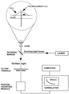
Figure 1
Figure 1
FCS experimental setup. Light from an argon ion laser is focused by means of a dichroic mirror and a lens to form a small volume element (0.2 fl). The laser beam is projected from below into a well containing a monolayer of cultured cells and tetramethylrhodamine (Rh)-labeled ligand (see magnified diagram at the top). After excitation of the labeled ligand, emitted light is transmitted via the dichroic mirror, a bandpass filter, and a pinhole to a photodetector. The volume element is positioned onto the cell surface with a microscope for detection of ligand binding. The dimensions of the laser beam focus and the pinhole together define the confocal volume element. The detector signal is fed into a digital signal correlator, which calculates the autocorrelation function of the detected intensity fluctuations.
Figure 2
Figure 2
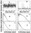
C-peptide binding and displacement to the membranes of cultured renal tubular cells. Fluorescence intensity fluctuations (A) and autocorrelation function (B) for Rh-CP (5 nM) free in solution, τ = 0.15 ms. Fluorescence intensity fluctuations (D) and autocorrelation function (E) for Rh-CP bound to membranes on the cell surface. Diffusion times (τ) and corresponding fractions (y): τ1 = 80 ms, y1 = 0.75; τ2 = 1 ms, y2 = 0.15; τ3 = 0.15 ms, y3 = 0.1. Autocorrelation functions of displacement of membrane bound Rh-CP by postincubation of a thousandfold molar excess of nonlabeled C-peptide (F) and nonlabeled C-terminal pentapeptide (C). The observed and calculated data points are completely overlapping (B, C, E, and F).
Figure 3
Figure 3
contin distributions of diffusion times Pi) of C-peptide binding and displacement to the membranes of cultured renal tubular cells. Rh-CP free in the incubation medium (A), binding of Rh-CP to the cell membranes (B), displacement of membrane-bound Rh-CP by incubation with a thousandfold molar excess of nonlabeled C-peptide (C), and inhibition of membrane binding of Rh-CP after pretreatment of the cells with pertussis toxin (D).
Figure 4
Figure 4
C-peptide binding curve. Binding of Rh-labeled C-peptide to cell membranes of renal tubular cells. Fractional saturation of the membrane-bound Rh-CP (y) as a function of the ligand concentration (L) in the binding medium. Each data point represents the mean of at least six measurements. The binding curve was simulated with Kass = 3.3 × 10⁹ M−1 and n = 1. Scatchard plot is shown as Inset.
Figure 5
Figure 5
Time course of displacement of Rh-CP by nonlabeled C-peptide. After incubation of cells with 5 nM Rh-CP for 60 min, 5 μM nonlabeled C-peptide was added, and FCS measurements were carried out at given time intervals. Each data point represents the mean of at least six measurements. Log scale for the binding displacement process is shown as Inset.

"VSports手机版" References

    1. Steiner D F, Oyer P E. Proc Natl Acad Sci USA. 1967;57:473–480. - PMC - PubMed
    1. Steiner D F, Cunningham D, Spigelman L, Aten B. Science. 1967;157:697–700. - PubMed
    1. Johansson B-L, Sjöberg S, Wahren J. Diabetologia. 1992;35:121–128. - PubMed
    1. Sjöquist M, Huang W, Johansson B-L. Kidney Int. 1998;54:758–764. - PubMed
    1. Johansson B-L, Linde B, Wahren J. Diabetologia. 1992;35:1151–1158. - PubMed

"VSports注册入口" Publication types