<b dropzone="i4zAjmqo"></b> Skip to main page content
U.S. flag

An official website of the United States government

Dot gov

The . gov means it’s official. Federal government websites often end in . gov or . mil. Before sharing sensitive information, make sure you’re on a federal government site VSports app下载. .

Https

The site is secure V体育官网. The https:// ensures that you are connecting to the official website and that any information you provide is encrypted and transmitted securely. .

. 1999 Oct 15;13(20):2658-69.
doi: 10.1101/gad.13.20.2658.

Disruption of the ARF-Mdm2-p53 tumor suppressor pathway in Myc-induced lymphomagenesis

Affiliations

Disruption of the ARF-Mdm2-p53 tumor suppressor pathway in Myc-induced lymphomagenesis

C M Eischen (V体育官网入口) et al. Genes Dev. .

VSports注册入口 - Abstract

Transgenic mice expressing the c-Myc oncogene driven by the immunoglobulin heavy chain enhancer (Emu) develop B-cell lymphoma and exhibit a mean survival time of approximately 6 months. The protracted latent period before the onset of frank disease likely reflects the ability of c-Myc to induce a p53-dependent apoptotic program that initially protects animals against tumor formation but is disabled when overtly malignant cells emerge. In cultured primary mouse embryo fibroblasts, c-Myc activates the p19(ARF)-Mdm2-p53 tumor suppressor pathway, enhancing p53-dependent apoptosis but ultimately selecting for surviving immortalized cells that have sustained either p53 mutation or biallelic ARF deletion VSports手机版. Here we report that p53 and ARF also potentiate Myc-induced apoptosis in primary pre-B-cell cultures, and that spontaneous inactivation of the ARF-Mdm2-p53 pathway occurs frequently in tumors arising in Emu-myc transgenic mice. Many Emu-myc lymphomas sustained either p53 (28%) or ARF (24%) loss of function, whereas Mdm2 levels were elevated in others. Its overexpression in some tumors lacking p53 function raises the possibility that Mdm2 can contribute to lymphomagenesis by interacting with other targets. Emu-myc transgenic mice hemizygous for ARF displayed accelerated disease (11-week mean survival), and 80% of these tumors lost the wild-type ARF allele. All ARF-null Emu-myc mice died of lymphoma within a few weeks of birth. About half of the tumors arising in ARF hemizygous or ARF nullizygous Emu-myc transgenic mice also overexpressed Mdm2. Therefore, Myc activation strongly selects for spontaneous inactivation of the ARF-Mdm2-p53 pathway in vivo, cancelling its protective checkpoint function and accelerating progression to malignancy. .


Figures (VSports注册入口)

Figure 1
Figure 1
ARF–Mdm2–p53 circuitry. Myc rapidly induces p19ARF expression, but without evidence that Myc transactivates the ARF promoter, its action may be indirect. p19ARF binds directly with Mdm2 to neutralize its function and triggers p53-dependent transcription. In turn, Mdm2 is a p53-responsive gene (pathway A) whose activation cancels the p53 response. Through ill-defined mechanisms, p53 regulates negatively both ARF (pathway B) and Myc (pathway C). However, it is conceivable that the feedback loops affecting ARF and Myc are one and the same.
Figure 2
Figure 2
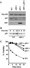
ARF and p53 mediate c-Myc-induced apoptosis in primary B cells. (A) Levels of Myc–ERTM, p53, and ARF in wild-type, ARF−/−, p53−/−, and ARF–p53 doubly null pre-B cells infected with a retrovirus encoding Myc–ERTM–GFP. Infected pre-B cells sorted for green fluorescence were expanded, lysed, and equal quantities of protein were assessed by immunoblotting with antibodies specific for each protein. (B) Kinetics of p53 induction in response to 4-HT in the same experiment shown in C. p53 was detected by immunoblotting as in A. No signals were detected in lysates of cells lacking p53 (negative data not shown). (C) Steady-state levels of apoptosis in the indicated primary pre-B cells at 14 days after infection are indicated at the 0 hr time point. 4-HT was added to the indicated primary pre-B cell cultures to activate Myc–ERTM, and their viability was determined at intervals thereafter by trypan blue dye exclusion and confirmed by analysis of subdiploid DNA content or staining with Hoescht 33342. The data are representative of three independent experiments.
Figure 3
Figure 3
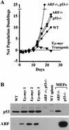
The ARF/p53 checkpoint is constitutively active in Eμ–myc transgenic bone marrow. (A) Growth curves of bone marrow cells explanted into pre-B cell culture medium containing IL-7. (B) Expression of p53 and p19ARF in explanted bone marrow cells at day 9 of culture. Pre-B cells were grown from the bone marrow of age- and sex-matched animals, including one nontransgenic wild-type (WT) mouse, three Eμ–myc transgenic mice, and from an animal lacking both ARF and p53. Spleen extract from a wild-type mouse and MEFs of the indicated genotypes were used as controls. Proteins were detected by immunoblotting as in Fig. 2.
Figure 4
Figure 4
p53, ARF, and Mdm2 expression and genotypic analysis of ARF and p53 in Eμ–myc lymphomas. (A) Levels of p53 (top), p19ARF (middle), and Mdm2 (bottom) in extracts of tumors from Eμ–myc transgenic mice were assessed by immunoblotting with antibodies specific for each protein. Extracts from ARF−/− and p53−/− MEFs (right two lanes) were used as controls. The asterisk in the left margin defines the position of a background band detected with Mdm2 C-18 antibody. Arrows indicate the locations of p53, p19ARF, and p92, p90, and p85 Mdm2 isoforms. (B) Southern blot analysis for genomic AflII and BamHI restriction fragments containing ARF exon1β (top) and p53 exons 2–10 (bottom), respectively. Tail DNAs extracted from ARF+/+, ARF+/−, and ARF−/− animals were used as controls (top, three right lanes) and indicate the positions of AflII restriction fragments containing (7.8 kb) or lacking (6.0 kb) ARF exon1β.
Figure 4
Figure 4
p53, ARF, and Mdm2 expression and genotypic analysis of ARF and p53 in Eμ–myc lymphomas. (A) Levels of p53 (top), p19ARF (middle), and Mdm2 (bottom) in extracts of tumors from Eμ–myc transgenic mice were assessed by immunoblotting with antibodies specific for each protein. Extracts from ARF−/− and p53−/− MEFs (right two lanes) were used as controls. The asterisk in the left margin defines the position of a background band detected with Mdm2 C-18 antibody. Arrows indicate the locations of p53, p19ARF, and p92, p90, and p85 Mdm2 isoforms. (B) Southern blot analysis for genomic AflII and BamHI restriction fragments containing ARF exon1β (top) and p53 exons 2–10 (bottom), respectively. Tail DNAs extracted from ARF+/+, ARF+/−, and ARF−/− animals were used as controls (top, three right lanes) and indicate the positions of AflII restriction fragments containing (7.8 kb) or lacking (6.0 kb) ARF exon1β.
Figure 5
Figure 5
Myc-induced tumorigenesis is accelerated by ARF loss. The genotypes of the mice are indicated next to the survival curves and the number of mice in each group are denoted by the n values. Thin vertical lines indicate ages of surviving mice. The average life spans of ARF+/+ Eμ–myc and ARF+/− Eμ–myc mice were 30 and 11 weeks, respectively. ARF−/− Eμ–myc animals were underrepresented because a fraction died soon after birth. Of those that survived weaning and were followed prospectively, their average survival was less than 7 weeks. Lymphoma was documented in all the animals.
Figure 6
Figure 6
ARF deletion and Mdm2 expression in tumors arising in ARF+/− and ARF−/− Eμ–myc mice. (A) Southern blot analysis of pre-B and B-cell tumors. Genomic DNAs digested with AflII were hybridized with a probe that detects a 7.8-kb fragment containing ARF exon1β and a mutant 6.0-kb fragment in which the exon was disrupted. The positions of fragments derived from the wild-type and mutant alleles are indicated at the left. Tail DNAs from wild-type, ARF+/−, or ARF null mice (lanes 1416) were run as controls. (B) Tumors from the same mice shown in A (with corresponding lane numbers) were analyzed for Mdm2 expression as in Fig. 4. Immunoblots were probed with Mdm2-specific antibody C-18 to detect p92, p90, and p85 isoforms. (C) Tumors from ARF−/− Eμ–myc animals were analyzed for Mdm2 expression as in B. Extracts of MEFs lacking both p53 and Mdm2 were used as a negative control.

References

    1. Adams JM, Cory S. Oncogene cooperation in leukaemogenesis. Cancer Surveys. 1992;15:119–141. - PubMed
    1. Adams JM, Harris AW, Pinkert CA, Corcoran LM, Alexander WS, Cory S, Palmiter RD, Brinster RL. The c-myc oncogene driven by immunoglobulin enhancers induces lymphoid malignancy in transgenic mice. Nature. 1985;318:533–538. - PubMed
    1. Alitalo K, Koskinen P, Makela TP, Saksela K, Sistonen L, Winqvist R. Myc oncogenes: Activation and amplification. Biochim Biophys Acta. 1987;907:1–32. - PubMed
    1. Alkema MJ, Jacobs H, van Lohuizen M, Berns A. Perturbation of B and T cell development and predisposition to lymphomagenesis in Eμ-bmi-1 transgenic mice require the Bmi-1 RING finger. Oncogene. 1997;15:899–910. - VSports最新版本 - PubMed
    1. Askew DS, Ashmun RA, Simmons BC, Cleveland JL. Constitutive c-myc expression in an IL3-dependent myeloid cell line suppresses cell cycle arrest and accelerates apoptosis. Oncogene. 1991;6:1915–1922. - PubMed

Publication types (VSports手机版)

V体育ios版 - MeSH terms

LinkOut - more resources