Skip to main page content
U.S. flag

An official website of the United States government

Dot gov

The . gov means it’s official. Federal government websites often end in VSports app下载. gov or . mil. Before sharing sensitive information, make sure you’re on a federal government site. .

Https

The site is secure. The https:// ensures that you are connecting to the official website and that any information you provide is encrypted and transmitted securely. V体育官网.

. 1999 Sep;19(9):5969-80.
doi: 10.1128/MCB.19.9.5969.

Impaired immune responses and B-cell proliferation in mice lacking the Id3 gene

Affiliations

Impaired immune responses and B-cell proliferation in mice lacking the Id3 gene (VSports最新版本)

L Pan et al. Mol Cell Biol. 1999 Sep.

Abstract

B-lymphocyte activation and proliferation induced by the B-cell receptor (BCR) signals are important steps in the initiation of humoral immune responses. How the BCR signals are translated by nuclear transcription factors into cell cycle progression is poorly understood. Id3 is an immediate-early gene responding to growth and mitogenic signals in many cell types including B cells. The primary function of the Id3 protein has been defined as that of inhibitor of basic-helix-loop-helix (bHLH) transcription factors. The interaction between Id3 and bHLH proteins, many of which are essential for cellular differentiation, has been proposed as a key regulatory event leading to cellular proliferation instead of differentiation. To further investigate the role of Id3 in tissue and embryo development and the mechanism of Id3-mediated growth regulation, we generated and analyzed Id3-deficient mice. While these mice display no overt abnormality in tissue and embryo development, their humoral immunity is compromised VSports手机版. The amounts of immunoglobulins produced in Id3-deficient mice immunized with a T-cell-dependent antigen and a type 2 T-cell-independent antigen are attenuated and severely impaired, respectively. Further analysis of lymphocytes isolated from Id3-deficient mice reveals a B-cell defect in their proliferation response to BCR cross-linking but not to lipopolysaccharide or a combination of BCR cross-linking and interleukin-4. Analyses of cultured lymphocytes also suggest involvement of Id3 in cytokine production in T cells and isotype switching in B cells. Finally, the proliferation defect in Id3-deficient B cells can be rescued by ectopic expression of Id1, a homologue of Id3. Taken together, these results define a necessary and specific role for Id3 in mediating signals from BCR to cell cycle progression during humoral immune responses. .


Figures

FIG. 1
FIG. 1
(A) Diagrams of the mouse Id3 genomic locus (top), the gene-targeting construct (middle), and the Id3 knockout allele (bottom). Exons and selection markers are indicated by solid and open boxes, respectively. The probe used for Southern analysis is shown. Restriction enzymes and selection markers are abbreviated as follows: B, BamHI; H, HindIII; P, PstI; S, SacI; X, XbaI; Xh, XhoI; PGK, phosphoglycerate kinase gene promoter; tk, thymidine kinase gene; neo, neomycin resistance gene. (B) Southern blot analysis of genomic DNA from Id3+/+, Id3+/−, and Id3−/− mice. DNA was digested with SacI and hybridized with the probe shown in panel A. The sizes of SacI fragments for wild-type and mutant alleles are 3.6 and 15 kb, respectively. (C) Northern blot analysis of RNA isolated from Id3+/+, Id3+/−, and Id3−/− mouse splenocytes. Probes used in hybridization are indicated on the left, with GAPDH as a loading control.
FIG. 2
FIG. 2
(A) T- and B-cell development in lymphoid tissues of Id3−/− mice. A three-color FACS analysis of lymphocytes from the thymus, spleen, bone marrow, and peritoneal cavity of Id3+/+ (left) and Id3−/− (right) mice is shown. Results are representative of multiple tests and are shown as two-color dot plots after eliminating the dead cells by 7AAD staining. The percentages of relevant lymphocyte population are indicated in the quadrants. (B) Surface marker expression and subpopulation of B cells in the spleens of Id3−/− mice. Splenocytes were isolated from Id3+/+ (left) and Id3−/− (right) mice and examined by flow cytometry analysis as described for panel A.
FIG. 3
FIG. 3
ELISA of Ig levels in serum of unimmunized Id3+/+ (n = 16) and Id3−/− (n = 16) mice. Results are presented as mean ± standard error for 16 mice for each genotype. Asterisks denote values that are significantly different from the wild-type controls (t test; P < 0.005).
FIG. 4
FIG. 4
(A) Humoral immune response to DNP-KLH. Id3+/+ (n = 5) and Id3−/− mice (n = 5) mice were injected i.p. with 100 μg of DNP-KLH in complete Freund’s adjuvant on day zero and were boosted without adjuvant 21 days later. The mice were bled at the indicated times for ELISA of DNP-specific antibodies. The relative concentrations of anti-DNP-specific antibody isotype are shown as mean optical density (O.D.) and standard error. Serum dilution factors are as follows: 1:1,000 for IgM and IgG1; 1:500 for IgG2a and IgG3; 1:100 for IgG2b and IgA. Id3−/− mice show significantly reduced IgG2a and IgG3 production on days 14 and 28 (t test against wild-type controls; P < 0.005). (B) Humoral immune response to DNP-Ficoll. Mice were injected i.p. with 25 μg of DNP-Ficoll on day zero and bled at the indicated times. DNP-specific antibodies for six different isotypes were analyzed by ELISA. Serum dilution factors are as follows: 1:1,000 for IgM and IgG3; 1:100 for the other four isotypes. A significant difference between the two genotypes was found in all cases following immunization (t test against wild type controls, n = 5 for each genotype; P < 0.005).
FIG. 5
FIG. 5
(A) Splenocytes were stained with FITC-labeled anti-B220 monoclonal antibody after being loaded with Indo-1AM. B220+ B cells were examined for [Ca2+]i following stimulation (indicated by arrows) with rabbit anti-mouse CD19 or F(ab′)2 goat anti-mouse IgM. [Ca2+]i was determined as the ratio of Indo-1 fluorescence at 405 nm to 525 nm. The results are representative of three pairs of Id3+/+ and Id3−/− mice. (B) Expression levels of MHC class II antigen (Ia) in splenic B cells purified from Id3+/+ (top) and Id3−/− (bottom) mice after a 24-h culture in the absence (dashed lines) or presence (solid lines) of 10 μg of F(ab′)2 goat anti-mouse IgM per ml.
FIG. 5
FIG. 5
(A) Splenocytes were stained with FITC-labeled anti-B220 monoclonal antibody after being loaded with Indo-1AM. B220+ B cells were examined for [Ca2+]i following stimulation (indicated by arrows) with rabbit anti-mouse CD19 or F(ab′)2 goat anti-mouse IgM. [Ca2+]i was determined as the ratio of Indo-1 fluorescence at 405 nm to 525 nm. The results are representative of three pairs of Id3+/+ and Id3−/− mice. (B) Expression levels of MHC class II antigen (Ia) in splenic B cells purified from Id3+/+ (top) and Id3−/− (bottom) mice after a 24-h culture in the absence (dashed lines) or presence (solid lines) of 10 μg of F(ab′)2 goat anti-mouse IgM per ml.
FIG. 6
FIG. 6
(A) B-cell proliferation induced by the F(ab′)2 fragment of goat anti-mouse IgM at three different concentrations (right) and by LPS (50 μg/ml), anti-CD40 monoclonal antibody (10 μg/ml), and 100 U of IL-4 per ml plus 5 μg of F(ab′)2 fragment of goat anti-mouse IgM per ml (left). Purified splenic B cells were cultured in the presence of the indicated stimuli for 64 h, with [3H]thymidine being added during the last 16 h. [3H]thymidine incorporation is shown as the mean and standard error of triplicate cultures. Results are representative of four independent experiments. (B) T-cell proliferation induced by anti-CD3 (10 μg/ml), ConA (2 μg/ml), and PMA (20 ng/ml) plus ionomycin (1 μg/ml) (P+I). Purified splenic T cells were cultured for 54 h, with [3H]thymidine being added during the last 16 h. Results are shown as in panel A and are representative of four independent experiments.
FIG. 6
FIG. 6
(A) B-cell proliferation induced by the F(ab′)2 fragment of goat anti-mouse IgM at three different concentrations (right) and by LPS (50 μg/ml), anti-CD40 monoclonal antibody (10 μg/ml), and 100 U of IL-4 per ml plus 5 μg of F(ab′)2 fragment of goat anti-mouse IgM per ml (left). Purified splenic B cells were cultured in the presence of the indicated stimuli for 64 h, with [3H]thymidine being added during the last 16 h. [3H]thymidine incorporation is shown as the mean and standard error of triplicate cultures. Results are representative of four independent experiments. (B) T-cell proliferation induced by anti-CD3 (10 μg/ml), ConA (2 μg/ml), and PMA (20 ng/ml) plus ionomycin (1 μg/ml) (P+I). Purified splenic T cells were cultured for 54 h, with [3H]thymidine being added during the last 16 h. Results are shown as in panel A and are representative of four independent experiments.
FIG. 7
FIG. 7
Induction of Id3 expression in purified splenic B cells stimulated with anti-IgM. RNA was isolated from the wild-type and Id3−/− mouse B cells stimulated with anti-IgM for the indicated times (0, 1.5, and 4 h). The expression of Id3, E2A, c-myc, and EF1α was determined by the RT-PCR assay with gene-specific primers. Each primer pair was tested for linear PCR amplification by running incremental PCR cycles. Results shown are the optimal cycle conditions for the given genes. Quantitation was further confirmed by repeating the entire proliferation and RNA analysis by using B-cells purified from a separate batch of animals.
FIG. 8
FIG. 8
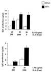
Ig secretion in stimulated splenic B cells. B cells from Id3+/+ and Id3−/− mice were purified from spleens and cultured for 6 days in the presence of various stimuli as indicated in the figure. The concentrations of Ig isotypes in the culture supernatants were determined by ELISA. Similar results were obtained from four independent experiments.
FIG. 9
FIG. 9
Cytokine gene expression in anti-CD3-stimulated Id3−/− mouse splenocytes. Erythrocyte-depleted splenocytes from Id3+/+ and Id3−/− mice were cultured with anti-CD3 for 48 h. Total RNAs were prepared, and cytokine gene expression was determined by an RNase protection assay. The identity of each band is indicated on the right. L32 and GAPDH are controls for mRNA quantity and quality.
FIG. 10
FIG. 10
(A) FACS analysis of bone marrow and splenic B cells from Id3+/+ (top) Id1tg (middle), and Id3−/− Id1tg (bottom) mice. The percentage of B220+/IgM+ cells is shown in the top right quadrant of each dot plot. (B) Comparison of Id3+/−, Id3+/− Id1tg, Id3−/−, and Id3−/− Id1tg mouse splenic B cells for their proliferative response to the F(ab′)2 fragment of anti-IgM (20 μg/ml), IgM (5 μg/ml) plus IL-4 (100 U), or LPS (100 μg/ml). [3H]thymidine incorporation is shown as mean and standard error of a triplicate culture. Results are representative of three independent experiments.

References

    1. Atherton G T, Travers H, Deed R, Norton J D. Regulation of cell differentiation in C2C12 myoblasts by the Id3 helix-loop-helix protein. Cell Growth Differ. 1996;7:1059–1066. - PubMed
    1. Bain G, Robanus Maandag E C, te Riele H P, Feeney A J, Sheehy A, Schlissel M, Shinton S A, Hardy R R, Murre C. Both E12 and E47 allow commitment to the B cell lineage. Immunity. 1997;6:145–154. - PubMed
    1. Bain G, Maandag E C, Izon D J, Amsen D, Kruisbeek A M, Weintraub B C, Krop I, Schlissel M S, Feeney A J, van Roon M, van der Valk M, te Riele H P J, Berns A, Murre C. E2A proteins are required for proper B cell development and initiation of immunoglobulin gene rearrangements. Cell. 1994;79:885–892. - PubMed
    1. Barone M V, Pepperkok R, Peverali F A, Philipson L. Id proteins control growth induction in mammalian cells. Proc Natl Acad Sci USA. 1994;91:4985–4988. - PMC (VSports最新版本) - PubMed
    1. Benezra R. An intermolecular disulfide bond stabilizes E2A homodimers and is required for DNA binding at physiological temperatures. Cell. 1994;79:1057–1067. - V体育官网入口 - PubMed

Publication types

MeSH terms

LinkOut - more resources