Skip to main page content
U.S. flag

An official website of the United States government

Dot gov

The . gov means it’s official. Federal government websites often end in VSports app下载. gov or . mil. Before sharing sensitive information, make sure you’re on a federal government site. .

Https

The site is secure. The https:// ensures that you are connecting to the official website and that any information you provide is encrypted and transmitted securely. V体育官网.

. 1999 Sep;73(9):7271-7.
doi: 10.1128/JVI.73.9.7271-7277.1999.

Inhibition of Epstein-Barr virus replication by a benzimidazole L-riboside: novel antiviral mechanism of 5, 6-dichloro-2-(isopropylamino)-1-beta-L-ribofuranosyl-1H-benzimidazole

Affiliations

V体育2025版 - Inhibition of Epstein-Barr virus replication by a benzimidazole L-riboside: novel antiviral mechanism of 5, 6-dichloro-2-(isopropylamino)-1-beta-L-ribofuranosyl-1H-benzimidazole

V L Zacny et al. J Virol. 1999 Sep.

Abstract

Although a number of antiviral drugs inhibit replication of Epstein-Barr virus (EBV) in cell culture, and acyclovir (ACV) suppresses replication in vivo, currently available drugs have not proven effective for treatment of EBV-associated diseases other than oral hairy leukoplakia. Benzimidazole riboside compounds represent a new class of antiviral compounds that are potent inhibitors of human cytomegalovirus (HCMV) replication but not of other herpesviruses. Here we characterize the effects of two compounds in this class against lytic replication of EBV induced in a Burkitt lymphoma cell line latently infected with EBV. We analyzed linear forms of EBV genomes, indicative of lytic replication, and episomal forms present in latently infected cells by terminal probe analysis followed by Southern blot hybridization as well as the high-molecular-weight unprocessed viral DNA by pulsed-field gel electrophoresis. D-Ribofuranosyl benzimidazole compounds that act as inhibitors of HCMV DNA maturation, including BDCRB (5, 6-dichloro-2-bromo-1-beta-D-ribofuranosyl-1H-benzimidazole), did not affect the accumulation of high-molecular-weight or monomeric forms of EBV DNA in the induced cells. In contrast, the generation of linear EBV DNA as well as precursor viral DNA was sensitive to the L-riboside 1263W94 [5, 6-dichloro-2-(isopropylamino)-1-beta-L-ribofuranosyl-1H-benzimidazole]. The 50% inhibitory concentration range for 1263W94 was 0. 15 to 1. 1 microM, compared with 10 microM for ACV. Thus, 1263W94 is a potent inhibitor of EBV. In addition, 1263W94 inhibited the phosphorylation and the accumulation of the essential EBV replicative cofactor, early antigen D VSports手机版. .


Figures

FIG. 1
FIG. 1
Structures of benzimidazole ribosides. BDCRB is the 2-substituted derivative of the compound DRB (5,6-dichloro-1-[β-d-ribofuranosyl) benzimidazole] (66, 67). The synthesis and activity of BDCRB have been described elsewhere (22, 68, 69). The compound 1263W94 differs from BDCRB in that it contains an l-sugar group and has a substitution of an isopropylamino group for bromine at the 2 position of the benzimidazole ring. Neither compound is phosphorylated in vitro or in vivo (5, 6).
FIG. 2
FIG. 2
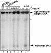
The l-riboside 1263W94 inhibits replication of EBV DNA. Induced Akata cells were incubated in the presence or absence of drug for 72 h and then harvested. Samples were prepared, and PFGE was performed to detect viral replication. EBV DNA was detected by hybridization with 32P-labeled EcoRI C fragment of EBV. HMW and monomer forms of DNA were quantitated following PhosphorImager analysis. A band representing nicked HMW DNA (∗) migrates to the resolution limit of the gel.
FIG. 3
FIG. 3
1263W94 inhibits replication of EBV. Akata cells were induced by IgG cross-linking and incubated in the presence or absence of drug for 3 days. Cells were harvested, and terminal probe analysis was performed as described in the text. DNA was digested with BamHI, separated on a 0.7% agarose gel, then transferred to nitrocellulose, and probed with 32P-labeled riboprobe Xho1.9 containing sequences specific for the EBV terminal repeat region. Samples containing ACV were incubated in the presence of TPase.
FIG. 4
FIG. 4
1263W94 inhibits appearance of EBV EA-D. Akata cells were induced by cross-linking with IgG and incubated in the presence or absence of 1263W94 for 3 days. Cells were harvested, and total protein lysate was prepared for immunoblot analysis; 30 μg of lysate were separated on an SDS–9% polyacrylamide gel, transferred to Immobilon, and probed with an EA-D-specific antibody. Detection was by enhanced chemiluminescence.
FIG. 5
FIG. 5
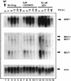
1263W94 does not inhibit the transcription of EA-D or the immediate-early genes R and Z. Akata cells were induced and then incubated in the absence of drug or with 5.0 μM 1263W94 or 50 μM ACV. Samples were harvested at times postinduction (p.i.), and total RNA was prepared. Northern blot analysis was performed with radiolabeled probes specific for EA-D, Z, or actin.
FIG. 6
FIG. 6
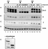
1263W94 inhibits the phosphorylation of EA-D. Akata cells were induced, and compound was immediately added. The samples were incubated for 24 h, then cycloheximide (CHX) was added to a final concentration of 50 μg/ml, and incubation was continued. Samples were harvested at the times indicated after addition of cycloheximide. Whole-cell lysates were prepared, and protein was quantitated by the Bradford method; 50 μg of each sample were separated on an SDS–10% polyacrylamide gel, and the protein was transferred to an Immobilon membrane. The membrane was probed with antibodies specific for EA-D (A), R (B), Z (C), or actin (D). (E) Akata cell lysates induced for 24 h (20 μg) were incubated with lambda phosphatase for 3 h at 30°C. The samples were separated by SDS-PAGE and then probed for EA-D as described above. un, untreated.
FIG. 7
FIG. 7
EA-D is underphosphorylated in DG75 cells. DG75 cells (107) were transfected with 5 μg of pSG5 vector (lane 1) or pSG5-BMRF1, with (lane 3) or without (lane 2) the addition of 5 μM 1263W94. Cells were harvested 24 h after transfection, and protein lysates were prepared. Akata cells (lanes 4 and 5) were induced by IgG cross-linking, and lysates were prepared from cells harvested 24 h postinduction. Following SDS-PAGE separation, the proteins were transferred and then probed with EA-D antibody as described in the text. Lane 5 is a shorter exposure of lane 4. Size at the left is indicated in kilodaltons.
FIG. 8
FIG. 8
EA-D phosphorylation is temporally regulated during the lytic cycle. Akata cells were induced by IgG cross-linking, and samples were harvested at 0, 12, 24, and 48 h postinduction (p.i.). Lysate samples (50 μg) were analyzed for EA-D protein.

"V体育官网" References

    1. Adams A, Lindahl T. Epstein-Barr virus genomes with properties of circular DNA molecules in carrier cells. Proc Natl Acad Sci USA. 1975;72:1477–1481. - PMC - PubMed
    1. Ben-Bassat H, Goldblum N, Mitrani S, Goldblum T, Yoffey J M, Cohen M M, Bentwich Z, Ramot B, Klein E, Klein G. Establishment in continuous culture of a new type of lymphocyte from a ‘Burkitt like’ malignant lymphoma (line DG-75) Int J Cancer. 1977;19:27–33. - PubMed
    1. Benz W C, Siegel P J, Baer J. Effects of adenine arabinoside on lymphocytes infected with Epstein-Barr virus. J Virol. 1978;27:475–482. - PMC - PubMed
    1. Biggen M, Bodescot M, Perricaudet M, Farrell P. Epstein-Barr virus gene expression in P3HR1-superinfected Raji cells. J Virol. 1987;61:3120–3122. - PMC - PubMed
    1. Biron, K. K. Unpublished data.

Publication types

MeSH terms