<acronym id="2PGNWdmf"></acronym>
Skip to main page content
U.S. flag

An official website of the United States government

Dot gov

The . gov means it’s official. Federal government websites often end in . gov or . mil. Before sharing sensitive information, make sure you’re on a federal government site VSports app下载. .

Https

The site is secure. The https:// ensures that you are connecting to the official website and that any information you provide is encrypted and transmitted securely. V体育官网.

. 1999 Apr 5;145(1):83-98.
doi: 10.1083/jcb.145.1.83.

GMAP-210, A cis-Golgi network-associated protein, is a minus end microtubule-binding protein (VSports最新版本)

Affiliations

"V体育2025版" GMAP-210, A cis-Golgi network-associated protein, is a minus end microtubule-binding protein

"V体育2025版" C Infante et al. J Cell Biol. .

Erratum in

  • J Cell Biol 2002 Aug 5;158(3):593

Abstract

We report that a peripheral Golgi protein with a molecular mass of 210 kD localized at the cis-Golgi network (Rios, R. M VSports手机版. , A. M. Tassin, C. Celati, C. Antony, M. C. Boissier, J. C. Homberg, and M. Bornens. 1994. J. Cell Biol. 125:997-1013) is a microtubule-binding protein that associates in situ with a subpopulation of stable microtubules. Interaction of this protein, now called GMAP-210, for Golgi microtubule-associated protein 210, with microtubules in vitro is direct, tight and nucleotide-independent. Biochemical analysis further suggests that GMAP-210 specifically binds to microtubule ends. The full-length cDNA encoding GMAP-210 predicts a protein of 1, 979 amino acids with a very long central coiled-coil domain. Deletion analyses in vitro show that the COOH terminus of GMAP-210 binds to microtubules whereas the NH2 terminus binds to Golgi membranes. Overexpression of GMAP-210-encoding cDNA induced a dramatic enlargement of the Golgi apparatus and perturbations in the microtubule network. These effects did not occur when a mutant lacking the COOH-terminal domain was expressed. When transfected in fusion with the green fluorescent protein, the NH2-terminal domain associated with the cis-Golgi network whereas the COOH-terminal microtubule-binding domain localized at the centrosome. Altogether these data support the view that GMAP-210 serves to link the cis-Golgi network to the minus ends of centrosome-nucleated microtubules. In addition, this interaction appears essential for ensuring the proper morphology and size of the Golgi apparatus. .


Figures
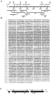
Figure 1
Figure 1
(A) Restriction map of the full-length GMAP-210 cDNA clone. Schematic representation of the cDNA clones obtained from a HeLa random-primed cDNA library (R1–R6, R8, and R9) and RT-7 and RT-10 clones obtained by RT-PCR. The full-length construct was assembled using clones R2, R3, R6, RT-7, and RT-10. The restriction map of GMAP-210 cDNA is represented at the top; the stippled region corresponds to the open reading frame. (B) Deduced amino acid sequence of GMAP-210. Regions predicted to form coiled-coil structures are in italics and boldface. Fragments used to generate polyclonal antibodies appear underlined. Proline residues disrupting coiled-coil are outlined. These sequence data are available from EMBL/GenBank under accession number Y12490. (C) Schematic representation of the secondary structure of GMAP-210. The data suggest an extended coiled-coil domain (boxes) interrupted by a non coiled-coil region of 100 amino acids and surrounded by a highly acidic NH2-terminal region containing a small coiled-coil domain and a basic proline/glycine-rich COOH terminus. Isoelectric points of the different regions are indicated at the top. LZ, leucine zipper; PP, proline-containing sequence.
Figure 8
Figure 8
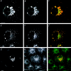
Expression of a GMAP-210 mutant form lacking the microtubule-binding site. After transfection, cells were fixed and double labeled for GMAP-210 (a) and the medial Golgi marker CTR433 (b). Red and green image pair is shown in c. Arrow in b indicates a nontransfected cell. Alternatively, cells were stained for GMAP-210 (d, e) and α-tubulin (f, g) and image pairs are shown in h and i, respectively. In d, f, and h, a transfected cell is shown. In e, g, and i, a nontransfected cell is presented for comparison. Bars, 10 μm.
Figure 12
Figure 12
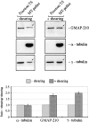
Binding of GMAP-210 to microtubules depends on the number rather than on the mass of microtubules. (Top) A NP-40–soluble and tubulin-free fraction was obtained from HeLa cells treated during 4 h with 20 μM taxol (fraction TS). This fraction contained a pool of GMAP-210 and γ-tubulin. Equal amounts of fraction TS were mixed with equal amounts of a suspension of taxol-induced microtubules from phosphocellulose-tubulin, before (−) or after (+) shearing through a 26-gauge hypodermic needle, and centrifuged through a sucrose cushion. Microtubule pellets were analyzed by IB. Shearing microtubules resulted in increased binding of γ-tubulin, indicating that more microtubule minus ends had been generated. Increased binding of GMAP-210 was also observed. (Bottom) Blots corresponding to three independent experiments were quantitatively analyzed using a Bio-Rad densitometer and NIH Image software. Quantification analysis indicates a twofold increase of bound γ-tubulin to sheared microtubules. In parallel, bound GMAP-210 increased 1.8 times.
Figure 2
Figure 2
Isolated cDNA codes for GMAP-210. Two polyclonal antibodies, named RM127 and RM130, were generated by immunizing rabbits with GST-Δ375-611 and GST-Δ618-803 fusion proteins. (A) These antibodies, the preimmune sera, and AS were assayed on blots of HeLa fractions. RM127 and RM130 revealed a band of identical electrophoretic mobility as that recognized by the AS that was absent from blots incubated with preimmune sera. (B) Endogenous GMAP-210 was immunoprecipitated from HeLa cells extracts using AS, RM127 or RM130 polyclonal antibodies linked to protein A–Sepharose beads. Immunoprecipitates were then analyzed by SDS-PAGE and IB with RM130 polyclonal antibody. Preimmune serum was included as a control.
Figure 7
Figure 7
Localization and effect of GMAP-210 overexpression on the Golgi apparatus and the microtubule network structure. Double IF was performed with RM130 (a, red) and CTR433 (b, green), or the AS (d, red) and anti-p115 (e, green). Separate red and green images were collected and overlaid (c and f). Yellowish staining indicates colocalization of GMAP-210 enriched membranes with the Golgi apparatus. Cells were also stained with RM130 (g, red) and an anti-tubulin antibody (h, green). Red and green images were collected separately and superposition of the two labelings is shown in i. Arrows in g and h indicate the Golgi apparatus and the microtubule aster in nontransfected cells. Bars, 10 μm.
Figure 9
Figure 9
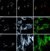
Effect of taxol on GMAP-210 overexpressing cells. Transfected cells were incubated with 10 μM taxol for 4 h and double stained for GMAP-210 (a, d, and g, red) and CTR433 (b, green) or tubulin (e and h, green). Superpositions of labelings are shown in c, f, and i. Preferential redistribution of GMAP-210–enriched membranes was observed at the cell periphery where the staining for GMAP-210 was equivalent to that observed in a juxtanuclear location (two opposed arrowheads in a). In b, the two unequal arrowheads indicate unequal partitioning of a Golgi marker that mostly remains in a juxtanuclear location. Note in f and i that GMAP-210–enriched Golgi elements are invariably associated with one end of microtubule bundles. Bars, 10 μm.
Figure 10
Figure 10
Transient expression of GFP-tagged NH2-terminal domain of GMAP-210. Cells were transfected with ΔC375-GFP (a, d, and g, green), processed and labeled with RM130 (b and e, red) or CTR433 (h, red). A nontransfected cell is shown in b and h for comparison. In c, f, and i superpositions of labelings are shown. Bars, 10 μm.
Figure 11
Figure 11
Transient expression of GFP-tagged COOH-terminal domain of GMAP-210. Cells were transfected with GFP-ΔN1778, processed and labeled with different markers. (a–c) A low magnification image showing a GFP-ΔN1778 expressing cell surrounded by several nontransfected cells (a, green). After transfection, cells were labeled with the centrosomal marker CTR453 (b, red). In c, superposition of both labelings is shown. Note that no staining with CTR453 could be detected in the GFP–COOH-terminal domain expressing cell. a1–3 (green) show increasing expression levels of GFP-ΔN1778; b1–3 (red) show staining with CTR453 and in c1–3 superpositions of both labelings are shown. (d–f) Effect of overexpression of GFP–COOH-terminal domain on the Golgi apparatus. Image pairs of GFP-ΔN1778 (green) and RM130 (d and e, red) or CTR433 (f, red) are presented. (g–i) GFP-ΔN1778 overexpressing cell (g, green) labeled for detyrosinated tubulin (h, red). Image pair is shown in i. Bars, 10 μm.
Figure 3
Figure 3
(A) Effects of microtubule-active drugs on the distribution of GMAP-210. HeLa cells were incubated with culture medium alone (Control), or with culture medium containing 10 μM nocodazole or 10 μM taxol for 4 h at 37°C. Cells were then extracted with 0.5% Triton in PHEM buffer for 5 min. Soluble (S) and insoluble (I) fractions were immunoblotted and blots probed with the AS, anti– α-tubulin antibody and anti-actin antibody as a control. (B) GMAP-210 binds to taxol-polymerized microtubules. HeLa cells were mechanically homogenized and the homogenates (H) were centrifuged at low speed to obtain a nuclear pellet (NP) and a post-nuclear supernatant (PNS). PNS was centrifuged at high speed and the supernatant (HSS) was incubated at 37°C for 15 min in the presence of 10 μM taxol. Microtubules (MTs) were sedimented through a cushion of 10% sucrose and all these subcellular fractions together with the microtubule-depleted supernatant (S) were then analyzed by SDS-PAGE and IB using AS and an anti–α-tubulin antibody. (C) Taxol-stabilized microtubules from HeLa cells were incubated with 0.5 M NaCl, 10 mM ATP, 10 mM GTP, and 2 M urea and equivalent amounts of supernatants (S) and pellets (P) analyzed by SDS-PAGE and IB for GMAP-210 and tubulin.
Figure 4
Figure 4
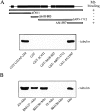
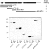
GMAP-210 binds directly to microtubules via its COOH-terminal domain. (A) Different portions of GMAP-210 or the complete protein were cloned in fusion with GST and immobilized on glutathione-agarose beads. Pure bovine brain tubulin was polymerized using taxol, microtubules were centrifuged through sucrose, washed and resuspended in PEM buffer. Polymerized microtubules were added to beads linked to GST alone, GST-fusion polypeptides, or GST-GMAP-210. After incubation and washing, the presence of microtubules in the bead pellets was revealed by IB using an anti–α-tubulin antibody. A summary of results obtained is shown at the top. The clones expressed as GST-fusions are represented as boxes. (B) Taxol-polymerized microtubules (lane Mts) were added to AS (lane AS+Mts), RM127 (lane RM127+Mts), and RM130 (lane RM130+Mts) immunoprecipitates from HeLa extracts, incubated and finally analyzed by SDS-PAGE and IB with anti–α-tubulin antibodies. As controls, preimmune serum was used instead of the immune sera (lane PI+Mts) or microtubules were not added (lane RM130−Mts).
Figure 5
Figure 5
GMAP-210 selectively interacts with a subpopulation of stable microtubules. (A) Untreated-, nocodazole- or taxol-treated HeLa cells were extracted for 5 min with 0.3% NP-40 in a microtubule-stabilizing buffer and the detergent-soluble fractions obtained were harvested (S fractions). Extracted cells were then incubated for 30 min on ice in the same buffer containing 5 mM CaCl2 in order to depolymerize microtubules (Ca fractions). All fractions together with the detergent- and calcium-insoluble fractions (I) were also analyzed by SDS-PAGE and IB using the AS and anti–α-tubulin antibodies. (B) HeLa cells grown on coverslips were treated and successively extracted with NP-40 and calcium as described in A. Cell ghosts were then processed for IF. The presence of microtubules was revealed by using an anti–α-tubulin antibody. Bars, 10 μm. (C) NP-40–soluble, CaCl2-soluble, and insoluble fractions from untreated HeLa cells were resolved by SDS-PAGE and blotted. Two identical blots were incubated with anti–α-tubulin antibody or with SG, a polyclonal antibody against detyrosinated tubulin (glu-tubulin). The same number of cells were used in each experiment, the different extractions were made in the same volume and identical volumes of samples analyzed in each line of gels. Blots were quantitatively analyzed using a Bio-Rad densitometer. Values are means of three independent experiments.
Figure 5
Figure 5
GMAP-210 selectively interacts with a subpopulation of stable microtubules. (A) Untreated-, nocodazole- or taxol-treated HeLa cells were extracted for 5 min with 0.3% NP-40 in a microtubule-stabilizing buffer and the detergent-soluble fractions obtained were harvested (S fractions). Extracted cells were then incubated for 30 min on ice in the same buffer containing 5 mM CaCl2 in order to depolymerize microtubules (Ca fractions). All fractions together with the detergent- and calcium-insoluble fractions (I) were also analyzed by SDS-PAGE and IB using the AS and anti–α-tubulin antibodies. (B) HeLa cells grown on coverslips were treated and successively extracted with NP-40 and calcium as described in A. Cell ghosts were then processed for IF. The presence of microtubules was revealed by using an anti–α-tubulin antibody. Bars, 10 μm. (C) NP-40–soluble, CaCl2-soluble, and insoluble fractions from untreated HeLa cells were resolved by SDS-PAGE and blotted. Two identical blots were incubated with anti–α-tubulin antibody or with SG, a polyclonal antibody against detyrosinated tubulin (glu-tubulin). The same number of cells were used in each experiment, the different extractions were made in the same volume and identical volumes of samples analyzed in each line of gels. Blots were quantitatively analyzed using a Bio-Rad densitometer. Values are means of three independent experiments.
Figure 6
Figure 6
NH2-terminal domain of GMAP-210 mediates its association to Golgi membranes. Different regions of GMAP-210 were expressed in fusion with GST and purified on glutathione-agarose beads. Beads were treated with thrombin to release recombinant polypeptides. Golgi membranes were purified by flotation in a sucrose gradient, washed with 2 M NaCl and centrifuged. 10 μg of Golgi membranes was incubated with <0.2 μg of recombinant polypeptides, centrifuged, and then supernatants and pellets were analyzed by IB using the AS. It must be noted that not all fragments were recognized with identical affinity by the AS. A summary of results obtained is shown at the top. The clones tested in the binding assay are represented as boxes.

References (V体育平台登录)

    1. Abe A, Emi NN, Tanimoto M, Terasaki H, Marunouchi T, Saito H. Fusion of the platelet-derived growth factor receptor β to a novel gene CEV14 in acute myelogenous leukemia after clonal evolution. Blood. 1997;90:4271–4277. - PubMed
    1. Ahmad FJ, Baas PW. Microtubules released from the neuronal centrosome are transported into the axon. J Cell Sci. 1995;108:2761–2769. - PubMed
    1. Alcalde J, Egea G, Sandoval IV. gp74, a membrane glycoprotein of the cis-Golgi network that cycles through the endoplasmic reticulum and intermediate compartment. J Cell Biol. 1994;124:649–665. - "V体育2025版" PMC - PubMed
    1. Bailly E, Doree M, Nurse P, Bornens M. p34cdc2 is located in both nucleus and cytoplasm; part is centrosomally associated at G2/M and enters vesicles at anaphase. EMBO (Eur Mol Biol Organ) J. 1989;8:3985–3995. - PMC - PubMed
    1. Bannykh SI, Balch WE. Membrane dynamics at the endoplasmic reticulum-Golgi interface. J Cell Biol. 1997;138:1–4. - PMC - PubMed

"VSports手机版" Publication types

MeSH terms