Skip to main page content
U.S. flag

An official website of the United States government

Dot gov

The . gov means it’s official. Federal government websites often end in . gov or . mil. Before sharing sensitive information, make sure you’re on a federal government site VSports app下载. .

Https

The site is secure. The https:// ensures that you are connecting to the official website and that any information you provide is encrypted and transmitted securely. V体育官网.

Comparative Study
. 1999 Apr;19(4):3145-55.
doi: 10.1128/MCB.19.4.3145.

SAG, a novel zinc RING finger protein that protects cells from apoptosis induced by redox agents (VSports最新版本)

Affiliations
Comparative Study

V体育ios版 - SAG, a novel zinc RING finger protein that protects cells from apoptosis induced by redox agents

H Duan et al. Mol Cell Biol. 1999 Apr.

Abstract

SAG (sensitive to apoptosis gene) was cloned as an inducible gene by 1,10-phenanthroline (OP), a redox-sensitive compound and an apoptosis inducer. SAG encodes a novel zinc RING finger protein that consists of 113 amino acids with a calculated molecular mass of 12. 6 kDa. SAG is highly conserved during evolution, with identities of 70% between human and Caenorhabditis elegans sequences and 55% between human and yeast sequences. In human tissues, SAG is ubiquitously expressed at high levels in skeletal muscles, heart, and testis. SAG is localized in both the cytoplasm and the nucleus of cells, and its gene was mapped to chromosome 3q22-24 VSports手机版. Bacterially expressed and purified human SAG binds to zinc and copper metal ions and prevents lipid peroxidation induced by copper or a free radical generator. When overexpressed in several human cell lines, SAG protects cells from apoptosis induced by redox agents (the metal chelator OP and zinc or copper metal ions). Mechanistically, SAG appears to inhibit and/or delay metal ion-induced cytochrome c release and caspase activation. Thus, SAG is a cellular protective molecule that appears to act as an antioxidant to inhibit apoptosis induced by metal ions and reactive oxygen species. .


VSports手机版 - Figures

FIG. 1
FIG. 1
SAG is evolutionarily conserved among different species. Primary amino acid sequences of human and mouse SAG were deduced from cDNAs cloned through DD and library screening. (A) Comparative alignments of SAG coding sequences from human, mouse, C. elegans, and yeast. Identity is shaded, and similarity is boxed. (B) Consensus sequence for the RING-H2 motif and comparison of the zinc RING finger domain of SAG with the RING-H2 motif. The C3H2C3 residues are in boldface.
FIG. 2
FIG. 2
SAG is inducible by OP. Subconfluent mouse L-RT101 and H-Tx cells were treated with OP (150 μM) for various periods of time up to 24 h and subjected to total RNA isolation and Northern analysis (with 15 μg of total RNA) using mouse SAG cDNA as a probe. Ethidium bromide staining of 28S and 18S rRNAs as loading controls is shown at the bottom.
FIG. 3
FIG. 3
SAG expression in multiple human tissues. hSAG cDNA was used as a probe for Northern analysis of poly(A)+ RNA isolated from different human tissues (Clontech). For an internal loading control, the housekeeping β-actin gene was used (bottom).
FIG. 4
FIG. 4
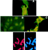
Cellular and chromosomal localization of human SAG. (A to C) Cellular localization. NIH 3T3 cells were plated onto coverslips, transiently transfected with plasmid encoding SAG-Myc tag, and immunofluoresced by antibody against Myc tag as detailed in Materials and Methods. Shown are the SAG-expressing cells (A), vector control cells (B), and LacZ-expressing cells (C). FISH mapping (bottom) was done as detailed in Materials and Methods. Shown are the FISH signals on the chromosome (left) and the same mitotic figure stained with DAPI to identify chromosome 3 (right).
FIG. 5
FIG. 5
SAG is a zinc binding protein. Shown are electrospray mass spectra of SAG protein in denaturing solvent (80:15:5 [vol/vol/vol] acetonitrile-water-acetic acid [pH 2.5]) (A) and nondenaturing solution (10 mM ammonium bicarbonate, 1 mM DTT [pH 7]) (B). The expected molecular mass of the apoprotein is 12,552 Da, with the first methionine deleted during expression and purification in bacteria. Assay conditions are detailed in Materials and Methods.
FIG. 6
FIG. 6
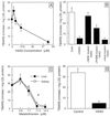
SAG inhibits LDL oxidation induced by copper or AAPH. (A) Dose-dependent inhibition of copper-induced LDL oxidation by SAG. Different amounts of SAG protein were incubated with LDL in the presence of CuSO4 (10 μM) for 10 min. Oxidation of LDL was measured by the formation of TBARS as detailed in Materials and Methods. (B) Abrogation of SAG protection by pretreatment with alkylating reagents but not by heat. SAG (750 μg/ml, 59 μM) was preincubated for 10 min with alkylating reagents N-ethylmaleimide (NEM; 50 mM) and p-hydroxymercuric benzoate (PHMB; 1 μmol/mg of SAG protein) or preheated (60°C for 15 min) before being subjected to LDL oxidation assay. Cont, control. (C) Dose-dependent inhibition of copper-induced LDL oxidation by MT. Different amounts of MT (from rabbit liver or horse kidney) were incubated with LDL in the presence of CuSO4 (10 μM) for 10 min. Oxidation of LDL was measured by the formation of TBARS. (D) Inhibition of LDL oxidation induced by AAPH. SAG (750 μg/ml, 59 μM) was incubated with LDL in the presence of AAPH (5 mM) for 10 min. Formation of TBARS was measured as a index of LDL oxidation.
FIG. 7
FIG. 7
Overexpression of hSAG protects DLD-1 colon carcinoma cells from apoptosis induced by OP and zinc. (A) Selection of SAG-expressing stable clones. DLD-1 cells were transfected with the neo control pcDNA3 or hSAG expression plasmid pcDNA-SAG. After G418 selection, resistant colonies were ring cloned and subjected to detection of exogenous SAG expression. (a) Expression of SAG mRNA. Total RNA was isolated and subjected to Northern analysis. Vector controls, D1-3 and D1-6; hSAG transfectants, D12-1 and D12-8. (b) 28S and 18S rRNAs for loading controls. (c) Expression of SAG protein in transfectants. The vector control lines and hSAG transfectants were subjected to immunoprecipitation. Shown is SAG protein expression in the neo controls (D1-3 and D1-6) and hSAG transfectants (D12-1 and D12-8). (B and C) hSAG overexpression protects cells from DNA fragmentation induced by OP or zinc. hSAG transfectants (D12-1 and D12-8), along with the vector control cells (D1-3 and D1-6), were seeded at 3.0 × 10⁶ to 3.5 × 10⁶ per 100-mm-diameter dish and exposed after 16 to 24 h to OP (150 μM; B) or zinc sulfate (125 μM; C) for 24 h. Both detached and attached cells in 2- by 100-mm dishes were harvested and subjected to DNA fragmentation assay. The 100-bp size marker is shown in the leftmost lane.
FIG. 8
FIG. 8
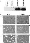
Overexpression of hSAG protects SY5Y neuroblastoma cells from apoptosis induced by copper and zinc. (A) Selection of SAG-expressing stable clones. SY5Y cells were transfected with the neo control pcDNA3 or hSAG expression plasmid pcDNA-Flag-SAG. After G418 selection, resistant cell lines were ring cloned and subjected to Western blot analysis for exogenous SAG expression with anti-Flag antibody. (B) Protection of metal-induced apoptosis as shown by morphological appearance. SAG-expressing cells (SYW20 [b, d, and f]) and neo control cells (SYV3 [a, c, and e]) were untreated (a and b) or exposed to CuSO4 (1.25 mM [c and d]) or ZnSO4 (200 μM [e and f]) for 16 h. Morphology was visualized at a magnification of ×200. (C) Protection of metal-induced apoptosis as shown by TUNEL assay. Cells were exposed to metal ions for 16 h as described above (without treatment [a and b]; with 1.25 mM CuSO4 [c and d]; with 200 μM ZnSO4 [e and f]), then subjected to TUNEL assay as detailed in Materials and Methods, and analyzed under a fluorescence microscope with a blue filter. Magnification, ×200.
FIG. 8
FIG. 8
Overexpression of hSAG protects SY5Y neuroblastoma cells from apoptosis induced by copper and zinc. (A) Selection of SAG-expressing stable clones. SY5Y cells were transfected with the neo control pcDNA3 or hSAG expression plasmid pcDNA-Flag-SAG. After G418 selection, resistant cell lines were ring cloned and subjected to Western blot analysis for exogenous SAG expression with anti-Flag antibody. (B) Protection of metal-induced apoptosis as shown by morphological appearance. SAG-expressing cells (SYW20 [b, d, and f]) and neo control cells (SYV3 [a, c, and e]) were untreated (a and b) or exposed to CuSO4 (1.25 mM [c and d]) or ZnSO4 (200 μM [e and f]) for 16 h. Morphology was visualized at a magnification of ×200. (C) Protection of metal-induced apoptosis as shown by TUNEL assay. Cells were exposed to metal ions for 16 h as described above (without treatment [a and b]; with 1.25 mM CuSO4 [c and d]; with 200 μM ZnSO4 [e and f]), then subjected to TUNEL assay as detailed in Materials and Methods, and analyzed under a fluorescence microscope with a blue filter. Magnification, ×200.
FIG. 9
FIG. 9
Overexpression of SAG inhibits or delays metal ion-induced cytochrome c release and caspase activation. (A) SAG-expressing DLD cells (D12-1) and neo control cells (D1-6) were subjected to ZnSO4 (140 μM) treatment for the indicated periods of time. The cytoplasmic fraction was extracted and subjected to Western blot analysis with antibodies against cytochrome c (top) and caspase 7 (bottom). (B) Human 293 cells were transiently transfected with hSAG expression plasmid or the neo control. Twenty-four hours posttransfection, cells were treated with CuSO4 (2 mM) for the indicated periods of time and subjected to Western blot analysis with antibodies against cytochrome c (top) and caspase 7 (bottom). Densitometric quantitation was performed in a densitometer. The band density from the untreated control was arbitrarily chosen as 1.

References

    1. Abello P A, Fidler S A, Bulkley G B, Buchman T G. Antioxidants modulate induction of programmed endothelial cell death (apoptosis) by endotoxin. Arch Surg. 1994;129:134–141. - PubMed
    1. Adebodun F, Post J F. Role of intracellular free Ca(II) and Zn(II) in dexamethasone-induced apoptosis and dexamethasone resistance in human leukemic CEM cell lines. J Cell Physiol. 1995;163:80–86. - VSports app下载 - PubMed
    1. Allen R G, Balin A K. Oxidative influence in development and differentiation: an overview of a free radical theory of development. Free Radical Biol Med. 1989;6:631–661. - PubMed
    1. Auld D S. Use of chelating agents to inhibit enzymes. Methods Enzymol. 1988;158:110–114. - "V体育安卓版" PubMed
    1. Baker A, Payne C M, Briehl M M, Powis G. Thioredoxin, a gene found overexpressed in human cancer, inhibits apoptosis in vitro and in vivo. Cancer Res. 1997;57:5162–5167. - PubMed

Publication types

MeSH terms

LinkOut - more resources